
Presentation
Female in 30's presents to A and E following a seizure.
Patient Data
Age: 30 years
From the case:
Sturge-Weber syndrome


Download
Info

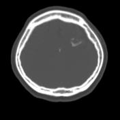
Gyriform calcification in the high left frontal lobe, with the characteristic 'tram-track' calcification.
Associated parenchyma volume loss.
Disproportionately large left frontal sinus compared to the right.
Choriod plexus symmeterical.
Case Discussion
Sturge Weber syndrome is one form of phakamatosis, characterized by a pial hemangioma in the brain with an associated facial hemangioma.
The leptomeningeal hemangioma results in a vascular steal affecting the subjacent cortex and white matter producing localized ischemia, hence the associated parenchymal volume loss.


 Unable to process the form. Check for errors and try again.