
Presentation
Acute onset of severe pain in the left knee. No history of trauma. The x-rays are not available.
Patient Data
Age: 60 years
Gender: Male
From the case:
Subchondral insufficiency fracture of the knee


Download
Info

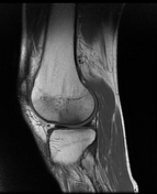
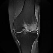
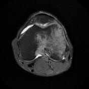
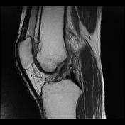
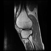
Subchondral lesion with mild irregularity and flattening of the inferocentral portion of the lateral femoral condyle (weight bearing surface). Extensive surrounding bone marrow edema extending to the adjacent soft tissues.
Moderate joint effusion is seen.
ACL, PCL, and collateral ligaments are intact. No meniscal tear is seen.
Case Discussion
The MRI features are most consistent with subchondral insufficiency fracture of the knee of the lateral femoral condyle, previously known as spontaneous osteonecrosis of the knee (SONK/SPONK) or Ahlbäck disease.


 Unable to process the form. Check for errors and try again.