
Presentation
Abdominal pain. Incidental finding.
Patient Data
Age: 30 years
Gender: Male
From the case:
Subhepatic cecum


Download
Info
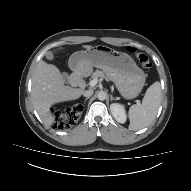
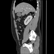
Diffusely decreased hepatic attenuation compared to the spleen in keeping with diffuse hepatic steatosis.
The cecum is of subhepatic location with a normal appearance of the terminal ileum and appendix.
Otherwise normal abdominopelvic CT exam.
From the case:
Subhepatic cecum
Download
Info

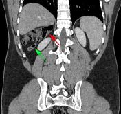
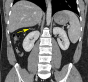
Annotated images:
red arrow = cecum
green arrow = terminal ileum
yellow arrow = appendix
Case Discussion
CT features of a subhepatic cecum which is due to the failure of the cecum to migrate to its typical position during midgut rotation in embryogenesis. Such a case may present a dilemma in patients with acute appendicitis where the pain or tenderness is located in the subhepatic region.


 Unable to process the form. Check for errors and try again.