
Presentation
Elevated D-dimer, palpitations.
Patient Data
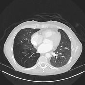

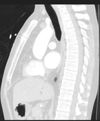
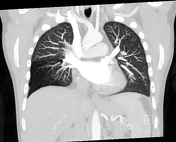
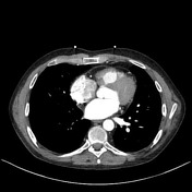

Mild bronchial wall thickening of the right lower lobe. Relative hyperlucency involving the majority of the right lower lobe and portions of the right middle lobe, with corresponding diminished size of the pulmonary vessels supplying these hyperlucent segments. Few other ill-defined areas of mosaicism in within the right upper lobe and left lower lobe.
Case Discussion
Swyer-James syndrome is post infectious obliterative bronchiolitis, usually a result of childhood infection. Commonly (on board examinations), it is described as "unilateral hyperlucent lung."
However, it often does not involve the entire lung, as in this case1. This is because the point of bronchial obstruction is in the small airways (bronchiolitis), not the larger segmental or central bronchi.
There is right lower and middle lobar hyperlucency, with a few scattered areas of mosaicism within the right upper lobe. There is corresponding diminished size of the pulmonary vasculature supplying the right middle and lower lobes. The left lower lobe has a few very faint areas of mosaic attenuation.


 Unable to process the form. Check for errors and try again.