
Presentation
Right thigh swelling.
Patient Data
Note: This case has been tagged as "legacy" as it no longer meets image preparation and/or other case publication guidelines.


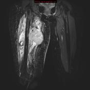
Rounded but ill-defined soft tissue swelling is located posteromedial to the femur. There is evidence of cortical erosion which appears to have a relatively wide zone of transition. The mass itself demonstrates no calcification. Incidental note is made of contrast in the bladder from the preceding CT scan (not shown).
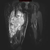
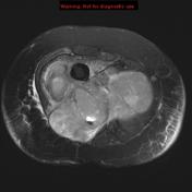

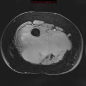
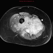
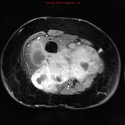
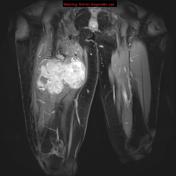

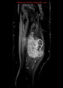

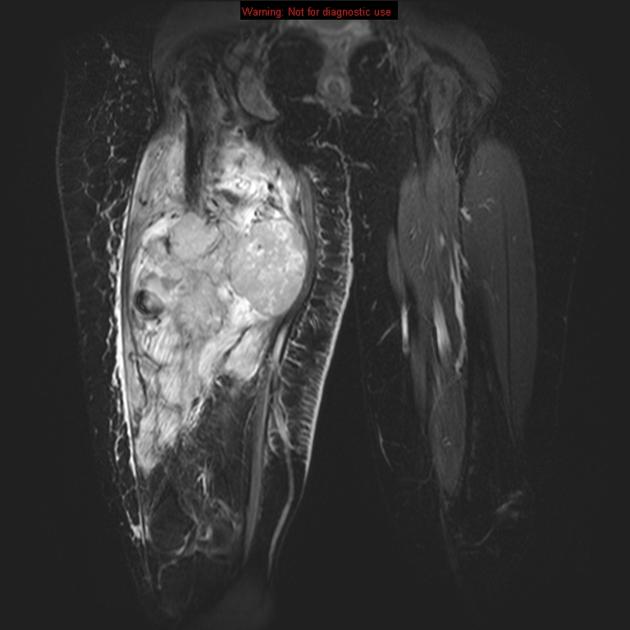
MRI of the thigh confirms the presence of a large heterogeneous mass extending into all compartments. It enhances brightly but irregularly with areas of presumed necrosis. Erosion of the femur is also shown without however evidence of extension into the bone marrow on axial images. There is extensive edema extending proximal and distal to the mass.

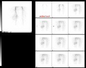
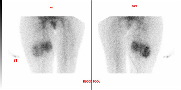
Bone scan demonstrates elevation of activity on both dynamic and blood pool views. Bone phase shows only minor local increase (not shown).
Case Discussion
Although synovial sarcomas are a relatively uncommon soft tissue sarcoma, they tend to affect younger patients and have a poor prognosis. They are difficult to distinguish from other sarcomas on imaging.


 Unable to process the form. Check for errors and try again.