
Presentation
25 year old female presenting with chest pain, left arm pain, parasthesia and headache. Clinical examination revealed diminished left upper limb pulsations. Echocardiography reveals mild pulmonary hypertension.
Patient Data
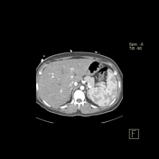


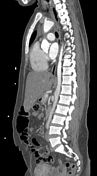
- diffuse thickening of the wall of entire thoracic and abdominal aorta with mild enhancement.
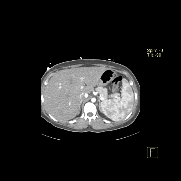
- diffuse long segment, very tight stenosis, of the scanned proximal left common carotid arteries as well as the left subclavian artery with enhancing thread like lumen.
- normal brachiocephalic artery as well as the right CCA and subclavian artery.
- moderate progressive attenuation of the abdominal aorta as well as the iliac arteries with decreased enhancement.
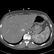
- the main pulmonary artery appears mildly dilated with diffuse mildly enhancing wall thickening of the pulmonary arteries. Mild diffuse attenuation of the left pulmonary artery as well as stenosis of the distal right pulmonary artery.
- no evidence of dissecting aortic flaps could be detected within the ascending or descending thoracic aorta. No evidence of periaortic leakage.
Case Discussion
The above described findings are those of Takayasu arteritis with diffuse aortitis, pulmonary arteritis, diffuse tight stenosis of the left CCA and subclavian artery, as well as stenosis of the left and right pulmonary arteries with mild pulmonary hypertension.
Takayasu arteritis (pulseless disease) is a chronic, granulomatous, large-vessel vasculitis affecting predominantly the aorta and its main branches in young females <30 years. Intimal proliferation eventually ends up with stenosis and occlusions of the affected arteries. Arbitrary classification of Takayasu arteritis includes 4 types:
Type 1: aortic arch and its main branches.
Type 2: abdominal aorta.
Type 3: entire aorta.
Type 4: pulmonary arteries are involved as well with peripheral pruning.
The main differential diagnosis is Giant cell arteritis. It is a vasculitis of medium-sized arteries; common in older patients >50 years. Aortitis (mostly the ascending aorta) in 10% of cases. Diagnosis is via biopsy of the superficial temporal artery.


 Unable to process the form. Check for errors and try again.