
From the case:
Talocalcaneal coalition
Download
Info
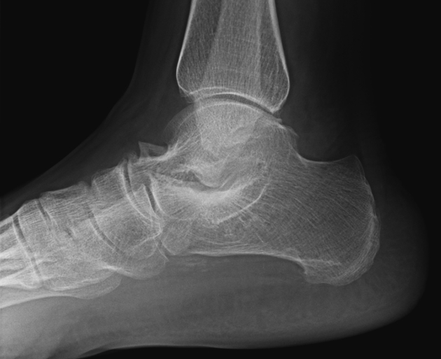
Talocalcaneal coalition. Note the talar beak and degenerative change at the talonavicular joint.
Case Discussion
Lateral view of the ankle demonstrates a talar beak or crest, a large and triangular flaring of the talar neck that ends at or near the articular margin of the talonavicular joint. More distally, an osteophyte can be seen at the talar head. In addition, there is obliteration of the posterior talocalcaneal joint, indicative of talocalcaneal coalition.
The talar beak should be differentiated from osteophytosis of the talar head (also seen in the image) and a normal or hypertrophied talar ridge.


 Unable to process the form. Check for errors and try again.