
Presentation
Chronic hindfoot pain.
Patient Data

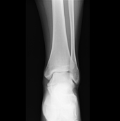
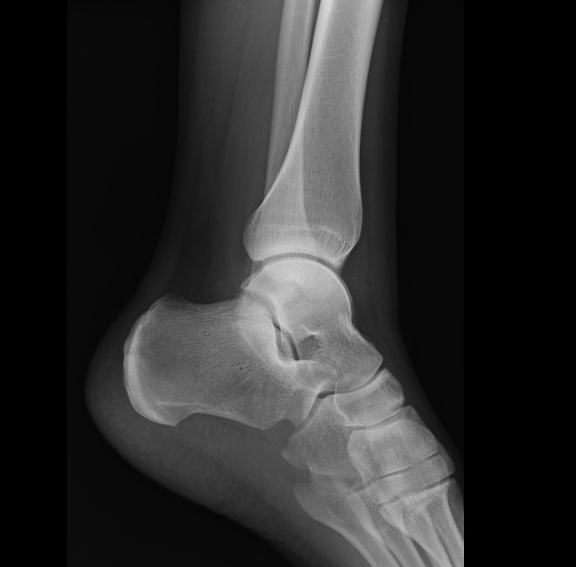
On the lateral x-ray, a C sign is present, suggestive of a talocalcaneal coalition. This is confirmed on a frontal view, where there is close apposition of a prominent medial talar dome and sustentaculum tali, indicating a non-osseous coalition.
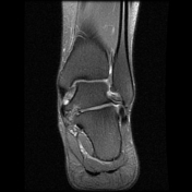

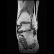
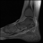
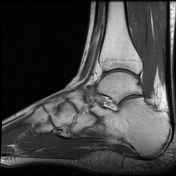
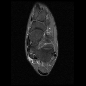

MRI confirms a non-osseous (cartilaginous) talocalcaneal coalition with secondary subchondral bony changes and bone marrow edema.
Case Discussion
Typical X-ray and MR findings of a talocalcaneal coalition are presented above.
The C sign helps detect the coalition on a lateral X-ray, but it can be present in both osseous and non-osseous types. In this case, a frontal view shows the lack of bony continuity in the area of interest, with abnormal articular orientation between the medial talar dome and the sustentaculum tali.
MRI aids in differentiating between types of the talocalcaneal coalition, including fibrous and cartilaginous, and can show reactive changes in adjacent bone and soft tissues. In this case, the signal between the medial talar dome and sustentaculum tali is similar to articular cartilage, indicative of the cartilaginous type.


 Unable to process the form. Check for errors and try again.