
Presentation
Chronic ankle pain with limitation of movement and no history of trauma
Patient Data
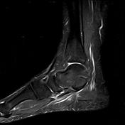

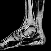


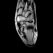
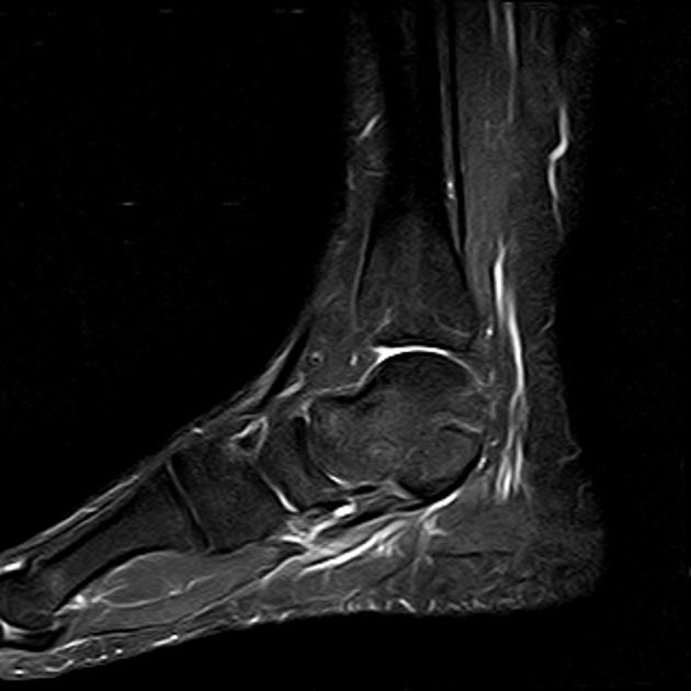
At the middle subtaral joint there is broadening of the talar facet and sustentaculum tali with reduced joint space and irregularities of the articular surfaces. Bone marrow edema is detected on STIR images. Also there is talar peak and pes planus deformity.
Diagnosis: talocalcaneal coalition - fibrous type


Features of talocalcaneal coalition including broadening of the middle subtalar joint articular surfaces (red circle), reduced joint space and irregularity of the articular surface with talar peak (orange circle)
Case Discussion
Tarsal coalition is abnormal fibrous/cartilagenous or bony fusion of two or more tarsal bones. Talar peak develops from excessive force on the talus from abnormal talocalcaneal articulation


 Unable to process the form. Check for errors and try again.