
Presentation
The patient has known thalassemia major. There is chronic growth retardation. MRI brain to exclude intracranial/ pituitary abnormality.
Patient Data


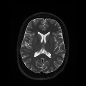
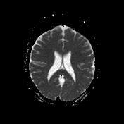
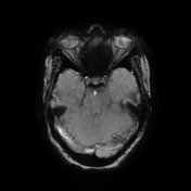
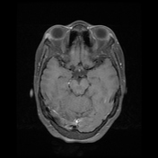
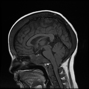

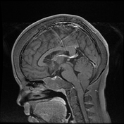
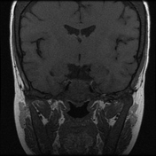
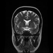
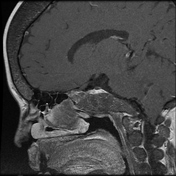
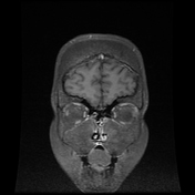
Multiparametric contrast-enhanced MRI brain demonstrates features of significant cranial medullary expansion. There is a widened diploic space, inner and outer cortical thinning, and sparing of the occipital bone.
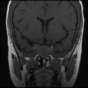
There is associated hypopneumatization of bilateral maxillary antra, sphenoids and frontal sinuses, replaced by red marrow. There is the expected sparing of the ethmoid sinuses.
There is a base of skull medullary expansion too.
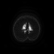
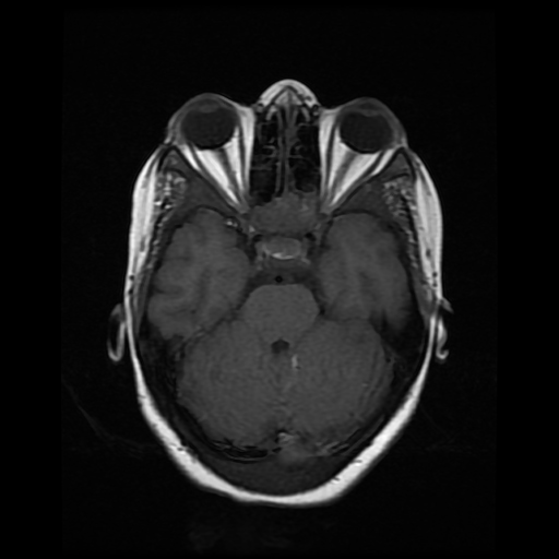
There is incidental Chiari I malformation (acquired), with cerebellar tonsillar herniation at 18.4mm. The visualized proximal cervical cord and brainstem appear normal.
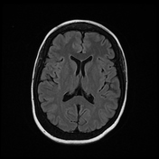
The pituitary gland is present and identified, measuring 9.9 x 3.5 x 5.3 mm (AP x cranio-caudal x width). The expected height of the pituitary gland for age should be 10 mm (patient 3.5 mm), features suggest possible pituitary hypoplasia in this setting. There is no pituitary macro- or microadenoma. There is no abnormal pituitary iron deposition in a setting of multiple blood transfusions and iron overload.
There is low T1 and T2 marrow signal within the proximal cervical spine due to marrow proliferation and conversion to red marrow.
Case Discussion
A known thalassemia major patient presents for exclusion of pituitary iron deposition and/or other intracranial pathology.
There are features consistent with the known history and consequent medullary expansion of the cranium and base of the skull. There is consequent development of inferior cerebellar tonsillar herniation and foraminal stenosis. The patient appears asymptomatic in this regard and has been referred for further clinical assessment with a view to a whole spine MRI. Based on the current imaging, features suggest an acquired Chiari I malformation.
There is a dramatic demonstration of the paranasal sinus hypopneumatisation and base of the skull medullary expansion.


 Unable to process the form. Check for errors and try again.