
Presentation
Abdominal pain and palpable pulsatile mass.
Patient Data


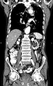
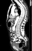

Ascending aorta is dilated up to 42 mm. Atherosclerotic changes and small pseudo aneurysm with mural thrombosis are observed at aortic arch. Descending aorta is also dilated particularly at distal portion measuring up to 50 mm containing mural thrombosis.
Abdominal aortic aneurysmal dilatation up to 78 mm is noted at infra renal segment contains marked mural thrombosis. Intraluminal aortic caliber measured about 38 mm. No peri aortic fluid or hematoma is noted.
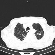
Fibrotic changes, focal calcifications, parenchymal distortion and tractional bronchiectasis are seen at both lung apices most compatible with sequel of old TB. Mild fibrotic changes are also observed at lower segments bilaterally.
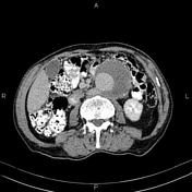
A few small calcified foci are seen at liver parenchyma most consistent with healed granuloma.
A 25 mm cortical cyst is present at upper pole of left kidney. Additionally, a 35 mm parapelvic cyst is noted at mid portion of the same kidney.
The prostate gland is enlarged.
Case Discussion
Multifocal thoracic and abdominal aortic aneurysm.


 Unable to process the form. Check for errors and try again.