
Presentation
Cough and dysphagia.
Patient Data
Age: 55 years
Gender: Female
From the case:
Thymoma
Download
Info
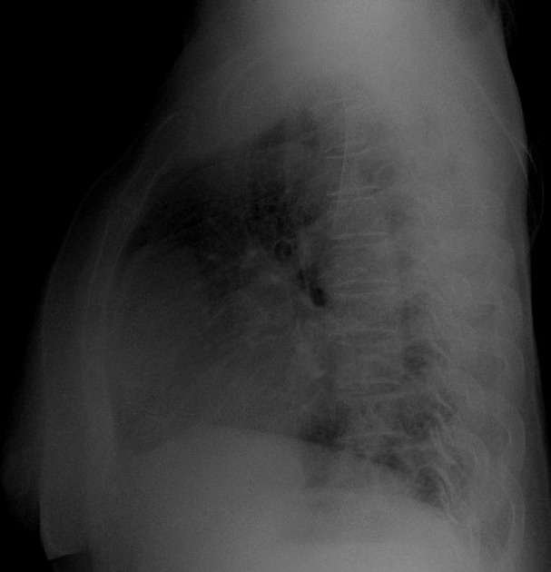
Frontal view shows a soft tissue shadow silhouetting the right heart border.
On lateral view an oval soft tissue shadow is appearing anterior to the heart.
From the case:
Thymoma


Download
Info

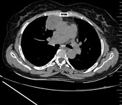
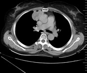
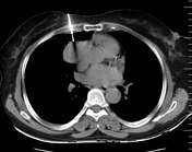
There is a well defined solid moderately enhancing lesion with a few non-enhancing necrotic areas within, is seen in the anterior mediastinum. No calcification cystic or fat component is seen within.
The lesion is anteriorly abutting the right mid costal cartilages and the sternum. However no obvious bony erosion or chest wall involvement is noted.
Postero-medially it is abutting the right atrium and the ascending aorta with maintained intervening fat planes, no infiltration to the adjacent structures is seen.
Imaging findings are suggestive of thymoma.
Biopsy was taken few days later and lesion turned out to be thymic epithelial hyperplasia.
Case Discussion
Thymoma is the most common primary tumor of anterior mediastinum and accounts for 20% of all mediastinal tumors. Although it can occur at any age, most patients are older than 40 years at the presentation. They are usually located anteriorly to the aortic arch but can occur in cardiophrenic angle.


 Unable to process the form. Check for errors and try again.