Presentation
Anterior neck swelling.
Patient Data
![](https://prod-images-static.radiopaedia.org/images/59227230/b75ff3f682ddbb9a9dc2d7821714357142691129f3c1725d0657113138f8b9cb_thumb.jpeg)
![This study is a stack](/packs/stack-YQKLCKBI.gif)
![](https://prod-images-static.radiopaedia.org/images/59227256/ef60ad78fb759a13e91bf608e24c0a679833281fab56cffbac16df382ee7be53_thumb.jpeg)
![This study is a stack](/packs/stack-YQKLCKBI.gif)
![](https://prod-images-static.radiopaedia.org/images/59227317/98b17ebeacd9eebfa93bc2306917124b464129ab4423cd54e286f629a85b5c3a_thumb.jpeg)
![This study is a stack](/packs/stack-YQKLCKBI.gif)
![](https://prod-images-static.radiopaedia.org/images/59227286/88bffc74f8bd0dea954e34433a27c18a9786630e88743998c95578b985a2e377_thumb.jpeg)
![This study is a stack](/packs/stack-YQKLCKBI.gif)
![](https://prod-images-static.radiopaedia.org/images/59227339/79089618d0be3bbad5cbad6310c6a44ee11ebb104df1f624f97b4ef0885b8c27_thumb.jpeg)
![This study is a stack](/packs/stack-YQKLCKBI.gif)
![](https://prod-images-static.radiopaedia.org/images/59227397/7cd72030b1f24d03cd96ff5149ee870efcc23b17ffade6fb942eb55506679328_thumb.jpeg)
![This study is a stack](/packs/stack-YQKLCKBI.gif)
![](https://prod-images-static.radiopaedia.org/images/59227358/dc4dbe1a8d254ee153297466a8c25dd38c7b73a6afb2f906d7452d016c5026fc_thumb.jpeg)
![This study is a stack](/packs/stack-YQKLCKBI.gif)
![](https://prod-images-static.radiopaedia.org/images/59227408/f21edad2e1c76608c95727d9b3e56a10c58875f57ba22b5ea6decc32c4984e27_thumb.jpeg)
![This study is a stack](/packs/stack-YQKLCKBI.gif)
![](https://prod-images-static.radiopaedia.org/images/59227230/b75ff3f682ddbb9a9dc2d7821714357142691129f3c1725d0657113138f8b9cb_big_gallery.jpeg)
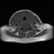
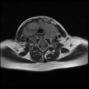
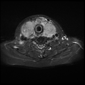
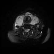
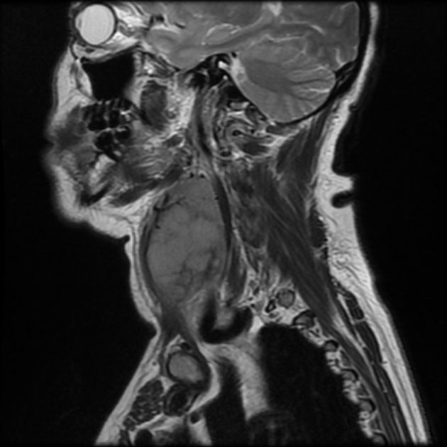
The thyroid gland is diffusely enlarged showing an abnormal increased signal on T2 and STIR. It shows diffusion restriction.
Multiple enlarged deep cervical, right retropharyngeal, pretracheal, and suprasternal lymph nodes.
![](https://prod-images-static.radiopaedia.org/images/59227443/cb9f48ea75ea5205921ad4b333dd4180e10fbc2f2fa5c5a6fe8b8d5058436e11_thumb.jpeg)
![This study is a stack](/packs/stack-YQKLCKBI.gif)
![](https://prod-images-static.radiopaedia.org/images/59227508/3266419f3403aa8a5dd7bac78507df7a0b1fae05b8a88d4b59778ca6293fc3bc_thumb.jpeg)
![This study is a stack](/packs/stack-YQKLCKBI.gif)
![](https://prod-images-static.radiopaedia.org/images/59227586/30d324b4d6d42fc008f02a1c1d156362bacacf1aea8ccfe0cb77799d570dbc12_thumb.jpeg)
![This study is a stack](/packs/stack-YQKLCKBI.gif)
![](https://prod-images-static.radiopaedia.org/images/59227652/30ad7a233e87b717a587cf08e0c644a986fb484117772db7c771664ba43cd7ed_thumb.jpeg)
![This study is a stack](/packs/stack-YQKLCKBI.gif)
![](https://prod-images-static.radiopaedia.org/images/59227443/cb9f48ea75ea5205921ad4b333dd4180e10fbc2f2fa5c5a6fe8b8d5058436e11_big_gallery.jpeg)
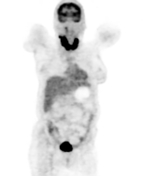
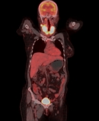
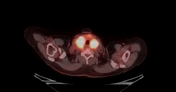
FDG avid diffusely enlarged thyroid gland with SUV max 15.
Multiple FDG avid right retrophayrngeal, deep cervical, pretracheal, and suprasternal lymph nodes.
US-guided biopsy (right thyroid lobe)
Gross description: Referred single H&E stained slide and 3 slides stained for antibodies (CD20, CD3, and cyclin D1).
Result:
The referred slides stained for:
CD20: positive: strong positive staining in lymphoid infiltrates
CD3: strong staining in reactive T lymphocytes
Cyclin D1: Negative in infiltrating lymphocytes
Performed immunohistochemical markers:
CD10: Negative
BCL6: Negative
BCL2: Negative
Conclusion:
Immunohistochemical features are consistent with extranodal marginal zone B-cell lymphoma.
Case Discussion
In this case, radiological criteria might suggest the possibility of thyroid lymphoma such as the diffuse involvement of the thyroid gland, the marked diffusion restriction, and the avid FDG uptake, as well as the distribution of the enlarged cervical lymph nodes.
Thyroid lymphoma is rare, accounting for a minority of both thyroid malignancies and lymphoma in general. The thyroid may be affected primarily or secondary to lymphoma elsewhere.
Hashimoto thyroiditis is a major risk factor (~60 times increased risk) but the development of thyroid lymphoma is still rare in this group.
Thyroid lymphoma may be a focal mass or diffuse gland enlargement.