
Presentation
Left tongue ulcer. Biopsy revealed invasive squamous cell carcinoma.
Patient Data
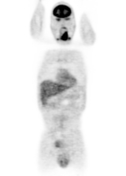

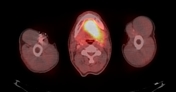
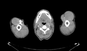
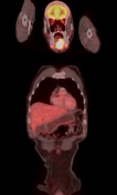
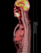
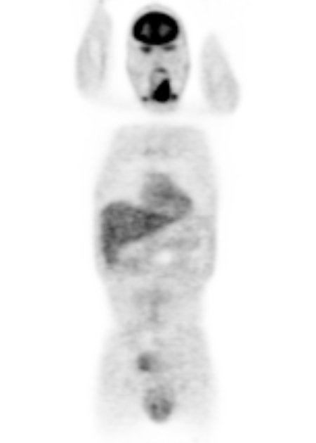
FDG avid soft tissue mass arising from the left side of tongue crossing the lingual septum and extending to the right para-midline region with SUV max 12.5 and measures 6.2 x 5 x 2.7 cm . The lesion invades the left genioglossus, geniohyoid, mylohyoid, and hyoglossus muscles. It infiltrates the hyoid bone anteriorly. It is also invading the left sublingual and submandibular salivary glands. The mass is abutting the adjacent mandible without erosion.
Lymph nodes: Increased FDG uptake by bilateral cervical lymph nodes level Ia, b, II, III with SUVmax 8.5 over 4 cm in left level II.
No distant metastasis.
Case Discussion
PET-CT is useful in staging tongue cancer, as in this case. It detected regional lymphadenopathy and excluded distant metastasis. However, MRI is better for local staging of tongue cancer as it better delineates the local invasion of the lesion.


 Unable to process the form. Check for errors and try again.