
Presentation
Hematuria.
Patient Data
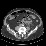

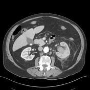
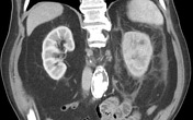
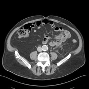
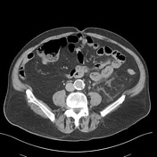

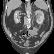
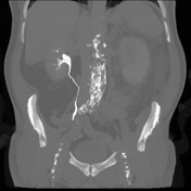

Hyperattenuation of the left renal collecting system extending into the left ureter with associated hydronephrosis and perinephric fat stranding and thickening of the perinephric fascia. Fullness within the upper pole collecting system measuring a maximum of 3 cm. Delayed cortical enhancement and excretion within the left kidney compared to the right side with failure to opacify the left renal collecting system at 10 min postcontrast administration. Normal contrast opacification of the right renal collecting system and ureter. No filling defect present within the right renal system. Foley catheter within the urinary bladder.
Gallstones within the gallbladder. The visualized spleen, visualized liver, adrenals, pancreas and small bowel are within normal limits. Diverticulosis of the sigmoid colon. Left para-aortic lymph nodes inferior to the left renal vein measuring short axis diameter of 1 cm. Ectatic abdominal aorta measuring a maximum of 3.3 centimeters.
Bibasal atelectasis. Spinal degenerative change. No suspicious osseous abnormality.
Impression:
Delayed left renal cortical enhancement with absence of contrast extravasation at 10 min consistent with impaired left renal function. Fullness of the left upper pole collecting system measuring a maximum of 3 cm with hyperattenuating debris within the left renal collecting system. CT findings are most consistent with an underlying mass and associated hemorrhage within the collecting system. Direct visualization and tissue sampling are recommended.
Case Discussion
Left nephro-ureterectomy revealed a TCC.


 Unable to process the form. Check for errors and try again.