
Presentation
Abdominal pain.
Patient Data
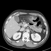

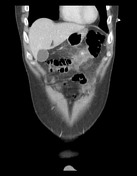

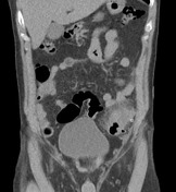
Short segment of wall thickening involving the mid portion of the transverse colon and associated with marked fat standing, particularly around a small anterior diverticular outpouching. Colonic diverticula identified scattered through the colon with no other foci of inflammatory changes. Bowel has otherwise unremarkable appearances. A small amount of free fluid is noted in the pelvis. No free gas in the peritoneal cavity. The liver, spleen, pancreas, kidneys and adrenal glands are normal. No suspicious lymph node enlargement. No suspicious bone lesions. Pleural bases are clear.

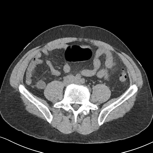
Short segment of abnormal circumferentially thickened colon at the junction of the descending colon into proximal sigmoid with regional diverticular the and extensive surrounding fat stranding with associated thickening of the adjacent parietal pleura. No adjacent collection or free gas identified. Small volume pelvic free fluid. No ureteric or pelvicalyceal calculi bilaterally. No hydronephrosis. Normal appendix. No abnormal small bowel dilatation.
Case Discussion
Case illustrating transverse colon diverticulitis in a patient that had presented 2 months earlier with similar symptoms due to a sigmoid colon diverticulitis.


 Unable to process the form. Check for errors and try again.