Presentation
Fall.
Patient Data
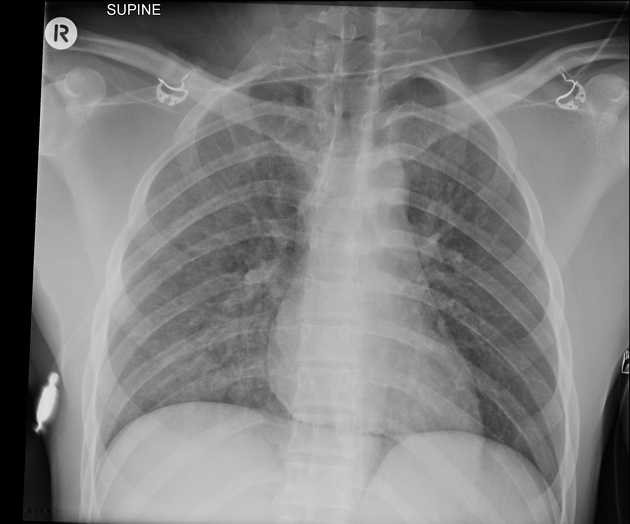
Supine trauma chest radiograph revealing subtle patchy opacity in the right hemithorax suspicious for pulmonary contusion. Subtle streaky linear lucency in the superior mediastinum and thin rim of air outlining the central portion of the diaphragm (continuous diaphragm sign) consistent with pneumomediastinum.
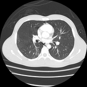

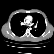

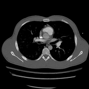

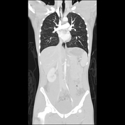

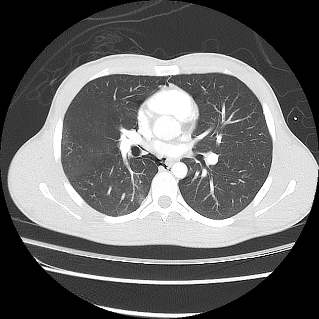
Right posterior pulmonary contusion particularly within the lower lobe. Associated right lower lobe pulmonary interstitial emphysema with air tracking into the mediastinum (pneumomediastinum). This mechanism of blunt traumatic pneumomediastiunum is known as the Macklin effect. Trace air below the heart in the epicardial fat explains the continuous diaphragm sign seen on x-ray. Small right pneumothorax predominantly anterobasally and trace left apical pneumothorax also present.
Case Discussion
Typical appearance of pulmonary contusion on CT with associated pneumomediastinum likely secondary to pulmonary interstitial emphysema (Macklin effect).


 Unable to process the form. Check for errors and try again.