
Presentation
The patient has eye contact abnormalities, delayed milestones.
Patient Data


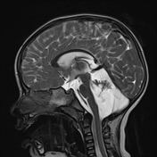
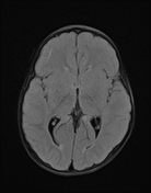
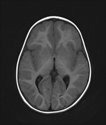
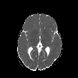
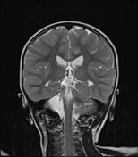
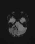
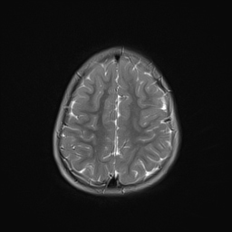
Absent (aplastic) right cerebellar hemisphere, right cerebellar vermis, and to a lesser extent right middle cerebellar peduncle.
An extra-axial large cystic lesion communicating with the fourth ventricle showing internal septa and ferromagnetic blooming artifact (likely due to hemosiderin deposition) is seen in place. It crosses the midline extending to the left side retro-cerebellar region.
Relatively thinned out corpus callosum (posterior portion of the body and splenium).
Relative flattening of the left posterior aspect of the midbrain.
Case Discussion
Cerebellar hypoplasia can be classified into two categories: focal and generalized. Focal hypoplasia is further divided into isolated vermian hypoplasia and hypoplasia of one cerebellar hemisphere.
Patients with unilateral cerebellar hypoplasia may be asymptomatic, or they may present with symptoms such as cerebellar dysfunction, headaches, or, in rare cases, seizures.
This condition's underlying cause may be a vascular insult that occurs during the intrauterine or perinatal period. As a result, there may be hypoplasia or aplasia of the cerebellar or vertebral arteries in these cases.


 Unable to process the form. Check for errors and try again.