
Presentation
Chronic left buttock pain.
Patient Data
Note: This case has been tagged as "legacy" as it no longer meets image preparation and/or other case publication guidelines.

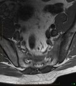
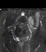

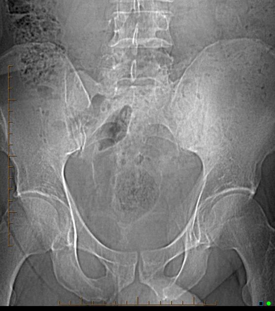
Left sacroiliac joint shows subarticular sclerosis surrounded by edema, appears to be inflammatory arthritis. No specific features to indicate an infective process and no ankylosis at this time. Right sacroiliac joint is normal.
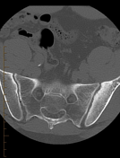
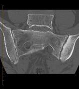
Unilateral left sacroiliac joint periarticular bone marrow sclerosis and articular erosions.
Case Discussion
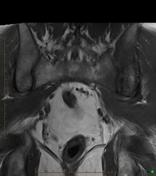
Unilateral periarticular bone marrow edema in the left sacroiliac joint with sclerosis and erosion on the CT scan. Appears to be inflammatory arthritis. No specific features to indicate an infective process and no ankylosis at this time. Right sacroiliac joint is normal on CT and MRI.


 Unable to process the form. Check for errors and try again.