
Presentation
Chronic pelvic pain and primary infertility.
Patient Data


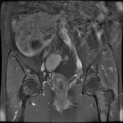
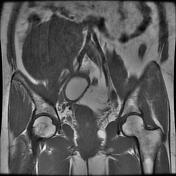
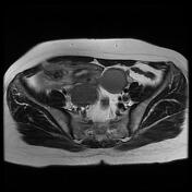
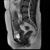
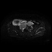
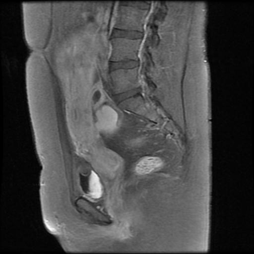
A well-defined large subserosal dominant myometrial lesion, appearing isointense on T1W and heterogeneous hypointense on T2W images, is seen arising from the right lateral wall and fundus of the uterus with intraabdominal extension above the level of the umbilicus, suggesting fibroid (Figo VI).
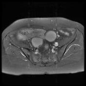
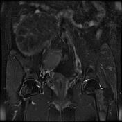
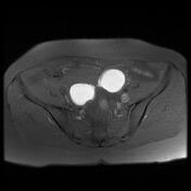
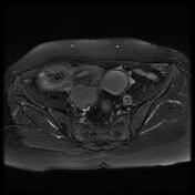
Two well-defined thin-walled cystic lesions are seen both ovary, appearing hyperintense on T1 fat-suppressed sequence, and hypointense on T2W images ('shading sign'), suggesting endometrioma cysts.
Both ovaries are seen in close proximity to each other behind the uterus (kissing ovaries).
Case Discussion
MRI study shows large subserosal uterine fibroid and bilateral ovarian endometrioma cysts (chocolate cysts) with T2 shading. The close proximity of both ovaries is known as "kissing ovaries" which is caused by adhesions and is considered a sign of pelvic endometriosis.


 Unable to process the form. Check for errors and try again.