
Presentation
Pelvic mass palpable on physical examination.
Patient Data
Age: 60 years
Gender: Female


Download
Info
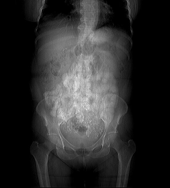
Scout image shows popcorn calcifications within the pelvis.
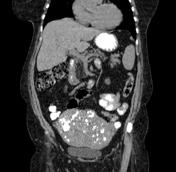
Coronal reformatting shows large uterine mass with lobulated margins and multiple coarse calcifications.
The patient also has a large known hepatic hemangioma (not adequately show in this sequence) and partial right ureteral duplication.
Case Discussion
On CT images, fibroids are usually of soft tissue density but may exhibit coarse peripheral or central calcification, as in this case. Enhancement pattern is variable.
The patient underwent a hysterectomy and the diagnosis was further confirmed on pathological examination.


 Unable to process the form. Check for errors and try again.