
Presentation
CT staging for breast cancer.
Patient Data
Age: 65 years
Gender: Female
Note: This case has been tagged as "legacy" as it no longer meets image preparation and/or other case publication guidelines.
Download
Info

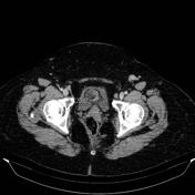
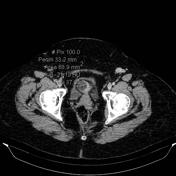
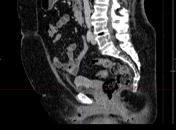
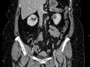
As an incidental finding is delimited a predominantly fat-containing well-demarcated myometrial nodule on uterine anterior body wall.
Case Discussion
This finding is already known and described in the prior ultrasound examination (not available).
Uterine lipoleiomyoma results from the degeneration of smooth muscle cells of an ordinary leiomyoma and represent a rare benign tumor of the uterus. It's often seen as a predominantly fat-containing well-demarcated mass with areas of soft tissue density arising from uterus, such as in this case.


 Unable to process the form. Check for errors and try again.