
Presentation
Pelvic pain
Patient Data
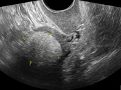

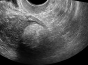
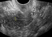

Ultrasound study shows left lateral uterine wall well-defined ovoid hyperechoic mass with a partially hypoechoic rim and posterior acoustic attenuation
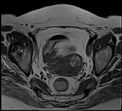

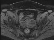
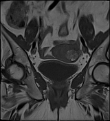
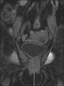
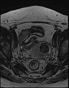
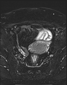
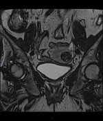
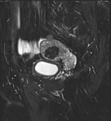

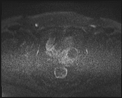
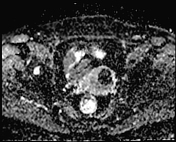
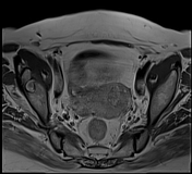
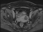
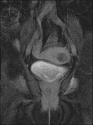

MRI study shows left lateral wall uterine well-defined ovoid slightly lobulated mass lesion centered upon the uterine myometrium measures 30 x 25 x 25 mm on its largest ap, tr, and cc dimensions respectively. It elicits heterogeneous signal intensity, with internal hyperintense areas on T1 and T2 that suppressed on T1 fat sat and STIR sequences respectively consistent with fatty contents. Minimal enhancement is noted on the post-contrast study.
No adnexal masses.
No pelvic collection.
Case Discussion
Uterine lipoleiomyomas result from degeneration of smooth muscle cells in an ordinary leiomyoma and represent a rare benign tumor of the uterus
Additional contribution from the radiologist; Dr Somia Elbadawy


 Unable to process the form. Check for errors and try again.