
Presentation
Asymptomatic; a hyperechoic uterine mass as an incidental finding on TVUS
Patient Data


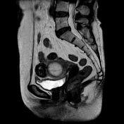
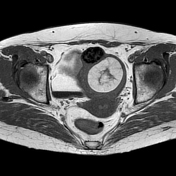
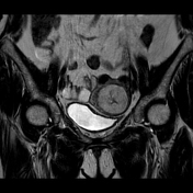
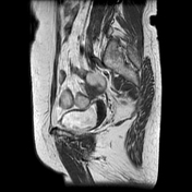
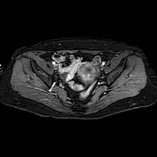
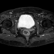
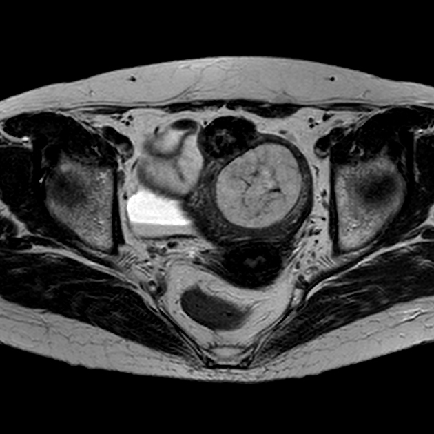
Solid intramural formation of the uterine body with well-circumscribed oval morphology that indents and thins the endometrial cavity. It is characterized by a predominantly hyperintense signal in T1 and T2 sequences, except for some thin linear low-signal bands likely attributable to smooth muscular or fibrotic tissue, hypointense in fat-sensitive sequences and without contrast enhancement. The finding is suggestive of a lipomatous uterine tumor, likely lipoleiomyoma.
Further smaller oval hypointense intramural and subserous masses are compatible with ordinary leiomyomas.
Accessory finding: urinary debris with layering in the bladder.
Case Discussion
Adipose degeneration of ordinary leiomyomas leads to the development of lipoleiomyomas, a benign and almost always asymptomatic finding. The detection of adipose tissue masses in the uterus is highly specific for diagnosis.


 Unable to process the form. Check for errors and try again.