
Presentation
Right neck swelling for 6 months.
Patient Data
Age: 25 years
Gender: Female


Show annotations
Download
Info
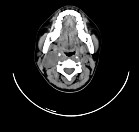
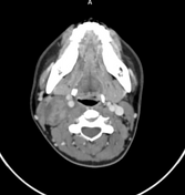
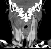
A well-defined oval-shaped soft tissue density lesion in the right upper cervical region below the sternocleidomastoid muscle. The lesion displaces the right internal & external carotid arteries medially and the internal jugular vein laterally. Mild patchy areas of heterogeneous enhancement were seen within the lesion on post-contrast images.
Case Discussion
The imaging findings are suggestive of vagal schwannoma.
The displacement of the internal carotid artery anteromedially and internal jugular vein laterally by the lesion is characteristic of vagal schwannoma.


 Unable to process the form. Check for errors and try again.