
Presentation
Abdominal distension
Patient Data


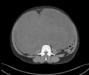
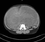
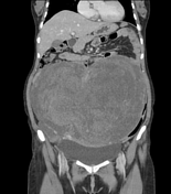
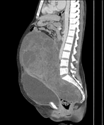
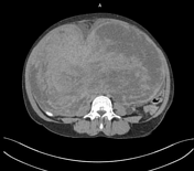
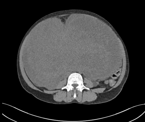
A large, well-defined, lobulated, heterogeneously enhancing mass lesion measuring approximately 32.8 x 28.5 x 17.4 cm is present in the lower abdomen and pelvis, extending up to the epigastric region. The lesion has both enhancing solid and non-enhancing cystic components, with marked internal vascularity in the solid component. Anteriorly, the lesion is causing displacement of the uterus with an ill-defined fat plane from the posterior wall of the uterus. No evidence of calcification or fat component was seen within the lesion. The bilateral ureters are displaced laterally by the mass lesion, with mild mass effect resulting in mild to moderate proximal hydroureteronephrosis on both sides. The mass lesion engulfs the right distal ureter. Both ovaries could not be delineated from the mass lesion. No ascites or locoregional lymphadenopathy was seen.
The findings are suggestive of a large solid-cystic abdominopelvic mass. Possible origins include ovarian or uterine.
Case Discussion
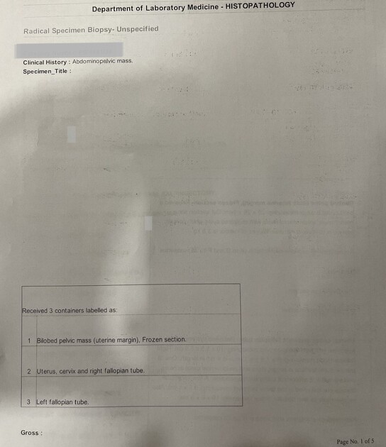
The patient was operated on, and the histopathological report of the radical specimen identified it as the vascular subtype of leiomyoma.
Co-author: Dr. Shweta Rai (senior consultant gynecologist).


 Unable to process the form. Check for errors and try again.