
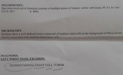
Presentation
Left wrist mass recurrence after two times of surgery.
Patient Data
Age: 30 years
Gender: Female
From the case:
Wrist tenosynovial giant cell tumor - recurrence

Download
Info

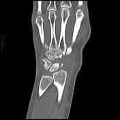
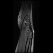
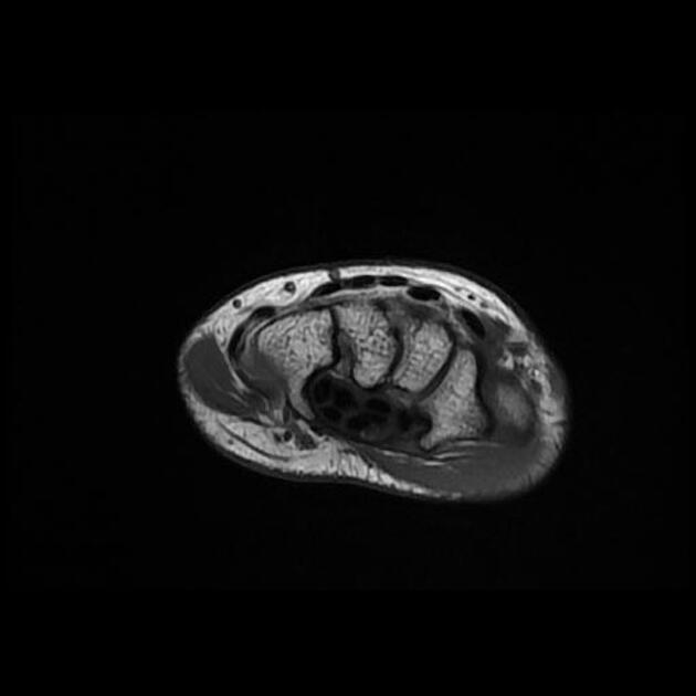
Soft tissue tumoral lesions are associated with well-defined lytic areas in the radius styloid, scaphoid, and triquetrum bones.
From the case:
Wrist tenosynovial giant cell tumor - recurrence


Download
Info
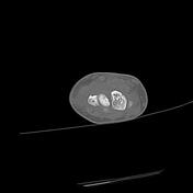
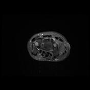
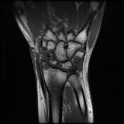
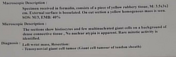
Abnormal signal enhancing soft tissue mass lesions penetrated into radius styloid, scaphoid, triquetrum bones, radiocarpal and intercarpal joints.
From the case:
Wrist tenosynovial giant cell tumor - recurrence
Download
Info
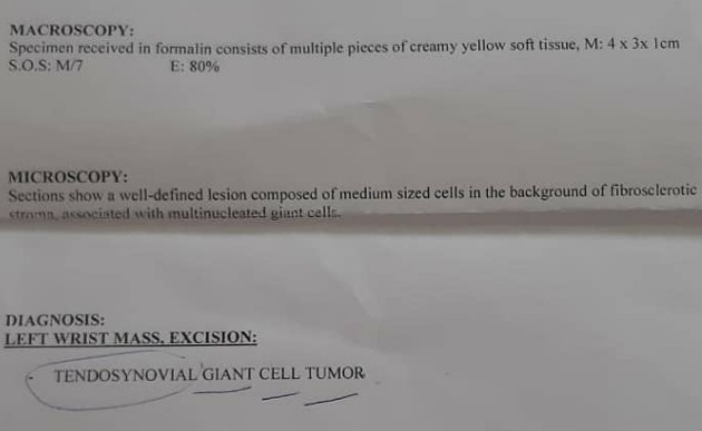
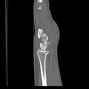
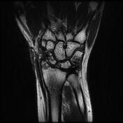
Tenosynovial giant cell tumor.
Case Discussion
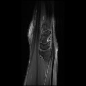
Imaging findings were compatible with recurrence/residue of the known wrist tumors (tenosynovial giant cell tumor).


 Unable to process the form. Check for errors and try again.