
Presentation
Vaginal bleeding.
Patient Data
Age: 9 months
Gender: Female
From the case:
Yolk sac tumor of infancy
Download
Info

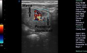
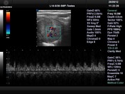
There is a lesion in the anterior myometriom that is hypervascular and which shows low resistance diastolic flow
From the case:
Yolk sac tumor of infancy


Download
Info
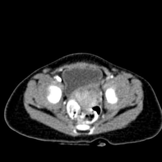
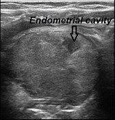
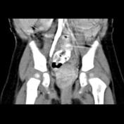
CT scan shows uterine enlargement which is definitely large for infants age.
From the case:
Yolk sac tumor of infancy


Download
Info
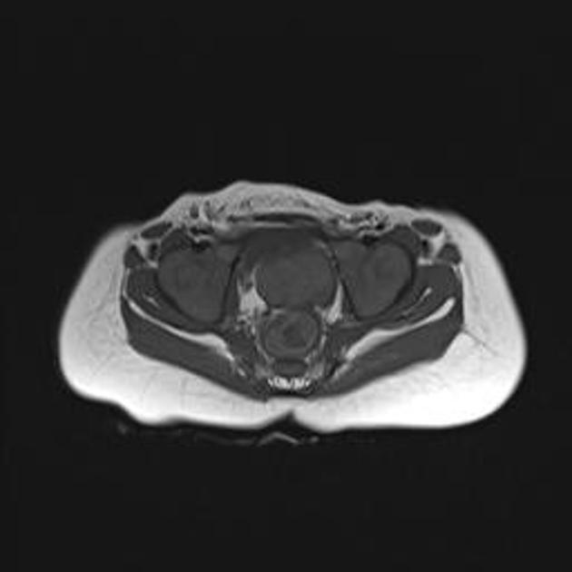
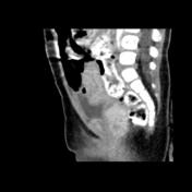
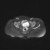
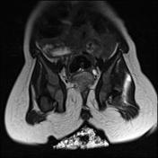
MRI shows a lobulated mass in the anatomic location of uterus which displays isointense signal on the T1 and slightly hypersignal on the T2 fat sat.
Case Discussion
The patient underwent surgery but the surgeon did not see a mass. Eventually vaginoscopy and biopsy was performed, proving a yolk sac tumor.


 Unable to process the form. Check for errors and try again.