BACK MENU NEXT

On this page:

Annotated images

Scroll to see annotations

Annotated image
This study is a stack

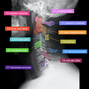
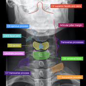
Case credit: Andrew Dixon, rID: 32505

Video presentation

Key Points

  • lateral radiograph
    • left and right facet joints should superimpose and appear like a single joint
      • except C2/3 facets which normally appear pseudo-fused 
    • mandibular angles should overlap - if not then there is lateral flexion or rotation
    • anterior and posterior tubercles of C1 should both look like the letter "D"
    • inferior vertebral body endplates are concave, while the superior endplates are flat
      • sharpened anterior inferior angle of the vertebral body is normal
    • transverse processes superimpose over the postero-superior corner of the vertebral body
  • frontal radiograph
    • airway creates a normal lucency over the vertebra
    • transverse processes:
      • C7 transverse process points horizontally or downward
      • T1 transverse process points superiorly
    • articular pillars form a sinusoidal wave along the lateral vertebral margin with the facet joint being the outward bulge

SKIP TO NEXT PAGE

Cervical spine series

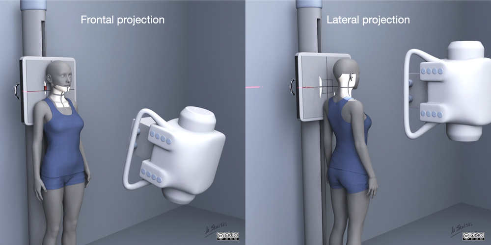
Illustration: Matt Skalski, rID: 80305

The cervical spine series is a set of radiographs taken to investigate the bony structures of the cervical spine, albeit commonly replaced by the CT, the cervical spine series is an essential trauma radiograph for all radiographers to understand.

Cervical spine radiographs are indicated for a variety of settings including 1-3:

  • trauma

  • infection

  • atypical pain

  • limb pain

  • osteoporosis

  • degenerative changes

A decision to pursue C-spine imaging of any kind should be cross-referenced with the 'Canadian C-Spine Rule' for C-spine imaging due to its high sensitivity and specificity 4.

Note: in the absence of CT 5 views of the C-spine should be performed: AP, lateral, obliques and odontoid 5.

  • AP  

    • anteroposterior projection of the cervical spine demonstrating the vertebral bodies and intervertebral spaces

  • lateral  

    • often utilized in trauma demonstrated

      • zygapophyseal joints 

      • soft tissue structures around the c spine 

      • spinous processes 

      • anterior-posterior relationship of the vertebral bodies 

  • odontoid  

  • AP oblique

    • demonstrates the intervertebral foramina of the side positioned further from the image receptor

  • PA oblique

    • demonstrated the intervertebral foramina of the side positioned closer to the image receptor

From article: Cervical spine series

Report problem MENU NEXT

:

Updating… Please wait.

 Unable to process the form. Check for errors and try again.

 Thank you for updating your details.