
Avulsion injuries or fractures occur where the joint capsule, ligament, tendon or muscle attachment site is pulled off from the bone, usually taking a fragment of cortical bone. Avulsion fractures are commonly distracted due to the high tensile forces involved. There are numerous sites at which these occur. Being familiar with them is important as subacute/chronic injuries can appear aggressive.
On this page:
Epidemiology
Avulsion injuries are common among those who participate in sports, in particular adolescents.
Pathology
Avulsion fractures can be classified as acute, subacute or chronic. In acute avulsion fractures, there is usually a clear preceding traumatic incident. Subacute and chronic avulsion injuries can be due to delayed presentation of an acute injury or secondary to repetitive use / overuse injuries 4.
The mechanism is from either 4:
high muscle activity
forced extreme range of motion
Location
Pectoral girdle
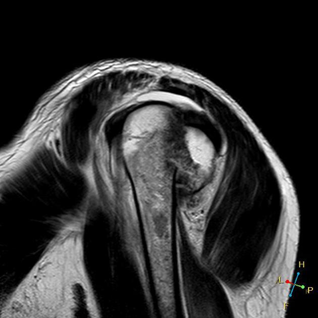
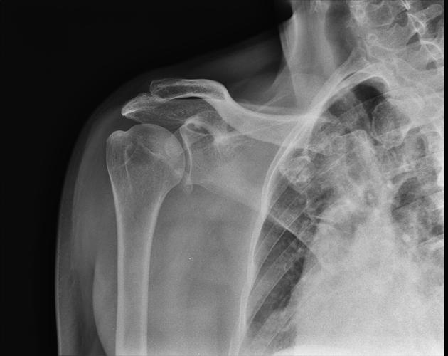
greater tuberosity: insertion of rotator cuff
lesser tuberosity: insertion of subscapularis (rare)
coracoclavicular avulsion
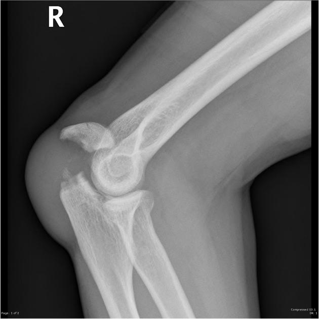
Elbow
-
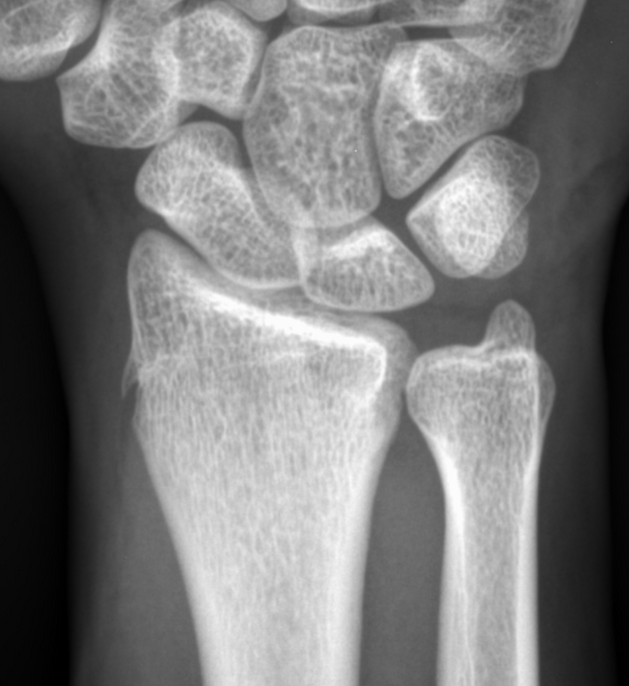
medial epicondyle: apophyseal avulsion in children
see also medial epicondylar fracture
coronoid process: insertion of capsule
radial tuberosity: insertion of biceps
olecranon process: insertion of triceps
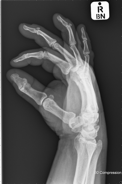
Hand
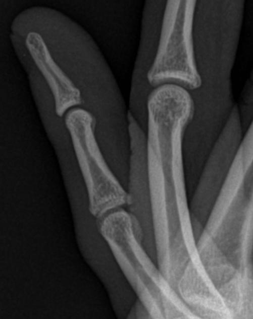
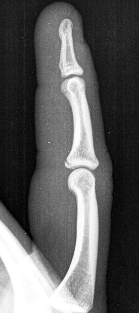
base of middle phalanx: volar plate avulsion injury
distal phalanx: mallet finger
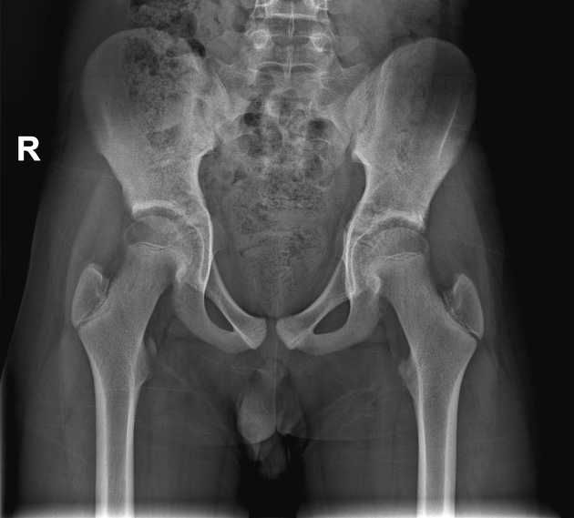
Pelvis and hip
iliac crest avulsion: anterior abdominal wall muscles
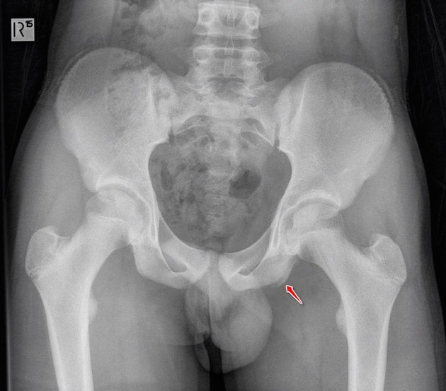
anterior superior iliac spine (ASIS) avulsion: tensor fascia lata and sartorius
anterior inferior iliac spine (AIIS) avulsion: straight head of rectus femoris
greater trochanter: hip rotator cuff
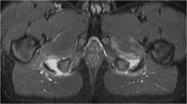
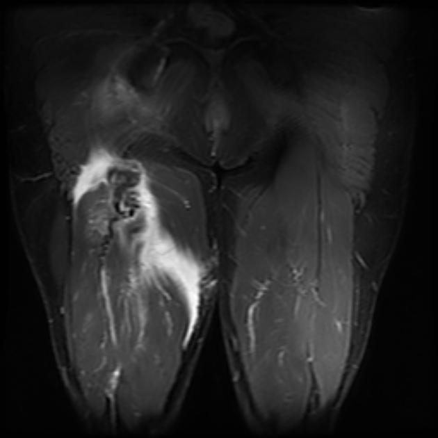
ischial tuberosity avulsion: hamstring muscles
body and inferior ramus of pubic bone: thigh adductors and gracilis
Knee
posterior tibial plateau/intercondylar area: posterior cruciate ligament
-
inferior pole of patella: patellar tendon
see also: Sinding-Larsen-Johansson syndrome and Jumper's knee
-
tibial tuberosity avulsion fracture: tibial tuberosity/patellar tendon
see also Osgood-Schlatter disease
lateral tibial plateau: lateral capsule
head of fibula: lateral collateral ligament and biceps femoris
-
medial aspect of femoral condyle: medial collateral ligament
see also: Pellegrini Stieda lesion
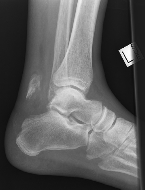
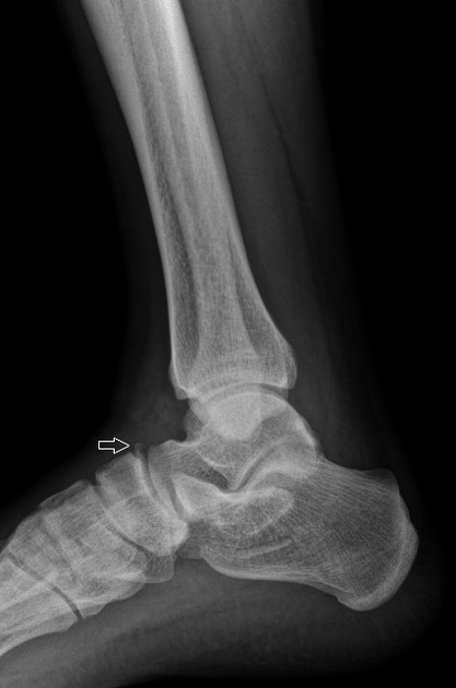
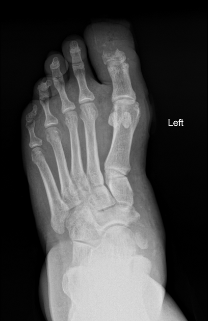
Ankle and foot
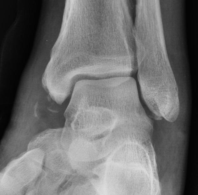
calcaneal tuberosity avulsion fracture: insertion of Achilles tendon
anterior process of the calcaneum: insertion of bifurcate ligament
dorsolateral process of the calcaneum: insertion of extensor digitorum brevis muscle
avulsion fracture 5th metatarsal styloid: insertion of peroneus brevis tendon
Radiographic features
Many avulsion fractures are apparent on plain radiographs. The avulsed bone fragment is typically displaced in the direction of the tendon, ligament or joint capsule which is attached to it 5. CT and/or MRI may be required for detection and further characterization. Appearances will vary depending on classification 4:
acute: avulsed bone fragment with donor site and typically associated soft tissue swelling / joint effusion
subacute: fracture healing results in a mixed lytic/sclerotic appearance
chronic: sclerosis and osseous hypertrophy
On MR small avulsion fractures can easily be missed, as the avulsed cortical fragment is often poorly visualized, and the bone marrow edema is absent at the site of injury 5.
Treatment and prognosis
Most avulsion injuries/fractures are treated non-operatively 4.
Differential diagnosis
accessory ossicle (some authors postulate that some accessory ossicles are the result from avulsion injuries, e.g. os subfibulare)
unfused ossification center


 Unable to process the form. Check for errors and try again.