
Anterior fontanelle inclusion cysts, also commonly referred to as congenital dermoid inclusion cysts or Adeloye-Odeku Disease are cystic lesions overlying the anterior fontanelle without communication with the underlying intracranial compartment.
On this page:
Terminology
Although the term congenital dermoid inclusion cyst is more common some of these lesions do not contain any secondary skin appendages and in fact represent epidermoid cysts 2. Furthermore, as even dermoids usually appear cystic without the fatty components frequently encountered elsewhere, it is difficult to distinguish between the two on imaging. As such anterior fontanelle inclusion cyst or anterior fontanelle dermoid/epidermoid is probably a better term.
Epidemiology
Congenital anterior fontanelle inclusion cysts are most frequently seen in black individuals or those with black ancestry and are more commonly seen in women (F:M 2:1) 2,3.
Clinical presentation
These cysts usually present at birth as rounded swellings over the anterior fontanelle without any pain or tenderness 2,3. Over time they gradually grow.
Occasionally these cysts can become infected 4.
Pathology
Anterior fontanelle inclusion cysts represent sequestration of epidermal rests during the third to fifth week of gestation 2. These cysts are lined by stratified squamous epithelium and their content primarily fluid 2. In the majority of cases, some secondary skin appendages (hair, sebaceous glands) are present and thus they represent dermoids. In others, only epidermal cells a found and thus these represent epidermoid cysts.
Radiographic features
The importance of imaging is to distinguish these cysts from other malformations that communicate with the intracranial compartment (see differential diagnosis section below). CT is, therefore, a minimum requirement, and if any concern for such communication exists, then further assessment with MRI is warranted.
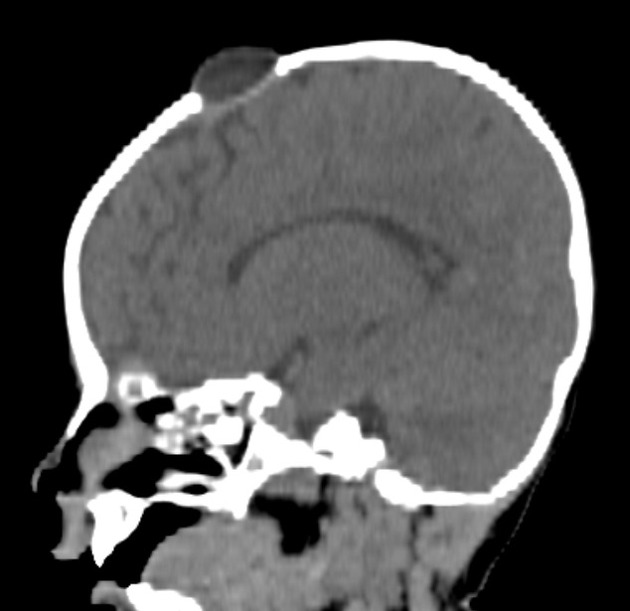
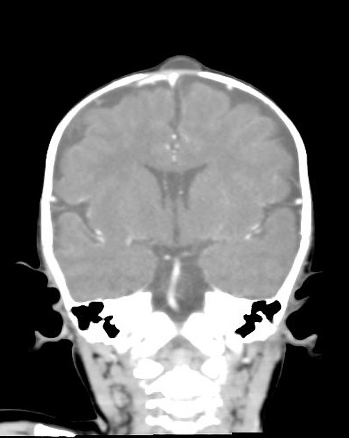
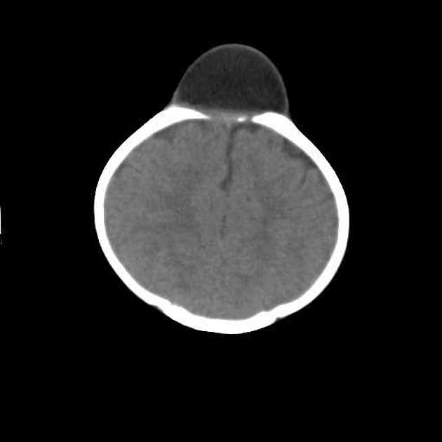
CT
These cysts appear as well-demarcated ovoid lesions ranging in size from a few millimetres to many centimetres in diameter. They are deep to the scalp and superficial to the dura. The fontanelle is often widened with remodelling/resorption of the underlying bone 4.
The content is hypodense similar to CSF but can be heterogeneous depending on content 4.
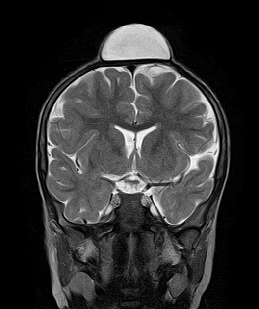
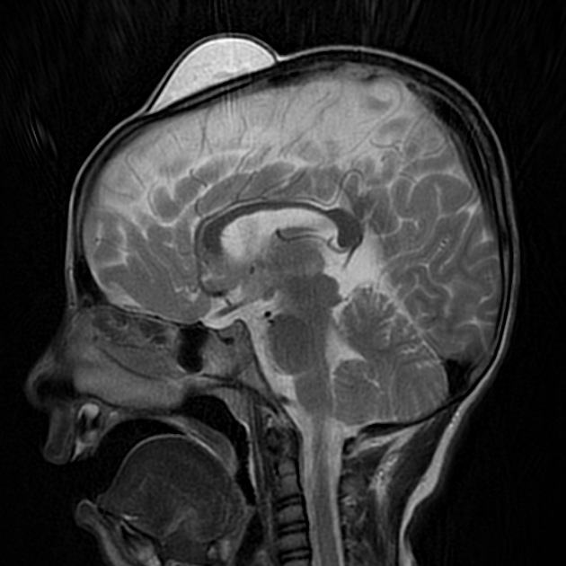
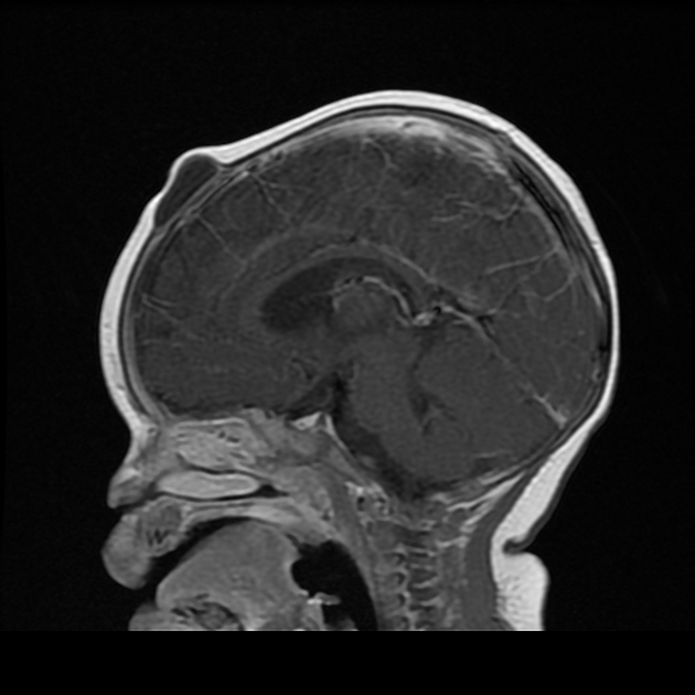
MRI
Most anterior fontanelle inclusion cysts are fluid-filled and therefore have low T1 and high T2 signal.
Treatment and prognosis
Surgical excision is necessary for cosmetic reasons and to confirm the diagnosis. Complete resection is curative.
History and etymology
It was first described by Adeloye and Odeku from Nigeria in 1971 1. It was considered a disease of African children. Other authors across the globe have subsequently reported, in various races, similar lesions.
Differential diagnosis
On imaging consider


 Unable to process the form. Check for errors and try again.