
Barrett oesophagus is a term for intestinal metaplasia of the oesophagus. It is considered the precursor lesion for oesophageal adenocarcinoma.
On this page:
Epidemiology
Barrett oesophagus is thought to have a prevalence of 3-15% in patients with reflux oesophagitis. Mean age at diagnosis is 55 years old 5.
Risk factors
Known risk factors for Barrett oesophagus include 10:
male gender
white race
-
~37% of patients (n = 27) who underwent upper endoscopy were found to have Barrett oesophagus 5
Clinical presentation
Barrett oesophagus is asymptomatic and usually discovered in a workup for GORD.
Pathology
Barrett oesophagus represents progressive metaplasia of oesophageal stratified squamous cell epithelium to columnar epithelium. Although the exact number varies, 90-100% of oesophageal adenocarcinoma is thought to arise from this metaplasia.
Although patients with Barrett oesophagus have a 30x risk of developing oesophageal adenocarcinoma 2, the annual risk of developing adenocarcinoma depends on the degree of histological dysplasia, but may be ~1% (range 0.1-2%), and the absolute risk is low 3.
Radiographic features
Because Barrett oesophagus represents metaplasia, it is often occult on imaging. Early oesophageal adenocarcinoma arising out of Barrett oesophagus also may be difficult to see. No radiological imaging modalities are adequate for screening.
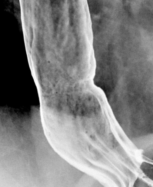
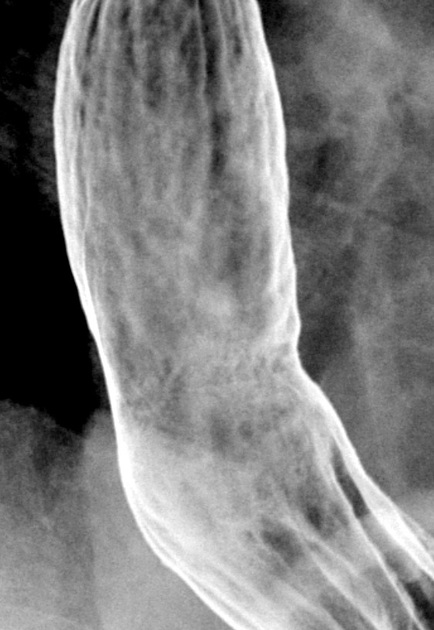
Fluoroscopy
-
double-contrast oesophagogram 7
-
signs of reflux oesophagitis
reflux
long stricture in the mid or lower oesophagus
large deep solitary ulcer
fine reticular mucosal pattern
thickened irregular mucosal folds
earliest signs of developing adenocarcinoma: localised flattening, stiffening, or irregularity in the wall of a stricture
-
There is a ~70% chance of Barrett oesophagus in a midthoracic oesophageal stricture ref.
Treatment and prognosis
Since Barrett oesophagus is considered a premalignant lesion, confirmation with upper endoscopy and biopsy is warranted.
If Barrett oesophagus is confirmed on biopsy, then aggressive therapy for gastro-oesophageal reflux is pursued, and perhaps endoscopic surveillance, depending on the patient's age and other risk factors.
One surveillance and biopsy protocol suggests 4:
low-grade dysplasia: 6-12 months
high-grade dysplasia: 3 months
If there is mucosal irregularity (what would be seen on an oesophagogram), then endoscopic resection has been recommended 4. Prophylactic resection or ablation has been used by some, particularly in younger patients.
History and etymology
Barrett oesophagus is named after Norman Rupert Barrett (1903-1979) 11, an Australian-born thoracic surgeon, who first described the condition in 1950 9.


 Unable to process the form. Check for errors and try again.