
Basilar artery aneurysms are less common than anterior circulation aneurysms, and rupture less frequently, but their critical location necessitates careful evaluation.
Unruptured basilar artery aneurysms occurs in 3% of all intracranial aneurysms 4.
Radiographic features
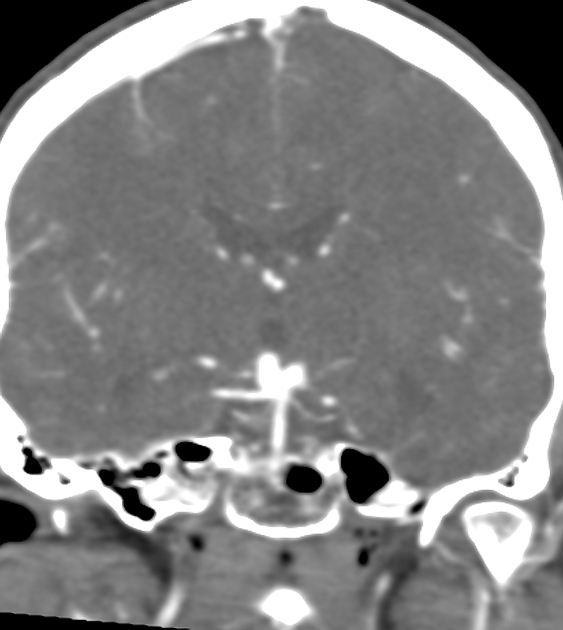
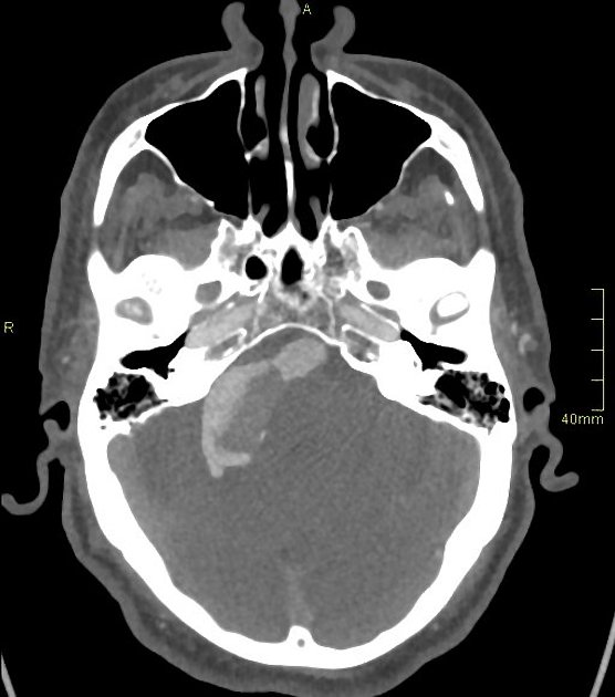
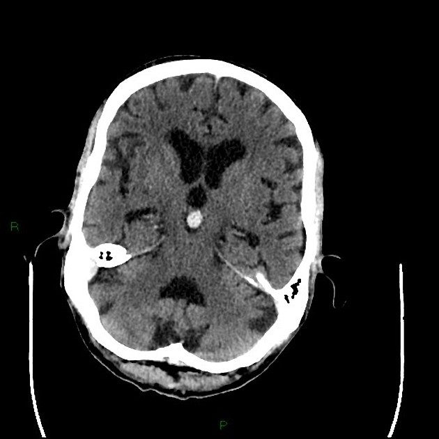
CT
may present as a lobulated hyperattenuating structure anterior to the mid brain
rupture of a basilar artery aneurysm is typically localised to the interpeduncular cistern, but may extend into the suprasellar cistern
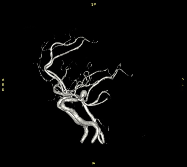
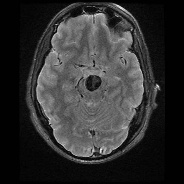
CT angiography (CTA) provides better evaluation of the aneurysm and its relationship to other branches of the basilar artery
basilar artery aneurysms can be both fusiform and saccular 2
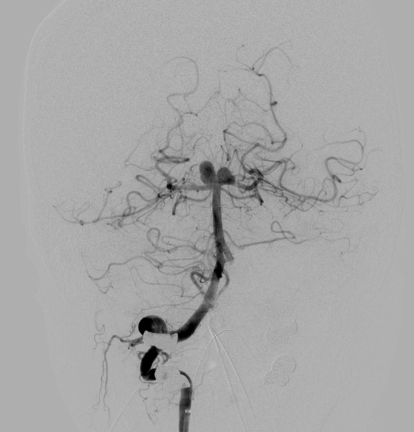
Angiography (DSA)
better than CTA for evaluating the relationship of the basilar aneurysm with branch vessels off the basilar artery, which is critical when considering intervention
Treatment and prognosis
Both unruptured and ruptured basilar artery aneurysms can be considered for clipping or endovascular coiling 4.
The type of treatment is tailored to the type of aneurysm (fusiform, saccular, branch, etc).
If coiled, they require close follow-up to ensure complete occlusion and may require re-treatment 3.


 Unable to process the form. Check for errors and try again.