Bathrocephaly
Citation, DOI, disclosures and article data
At the time the article was created Antonio Rodrigues de Aguiar Neto had no recorded disclosures.
View Antonio Rodrigues de Aguiar Neto's current disclosuresAt the time the article was last revised Arlene Campos had no financial relationships to ineligible companies to disclose.
View Arlene Campos's current disclosures- Bathrocephalic occiputs
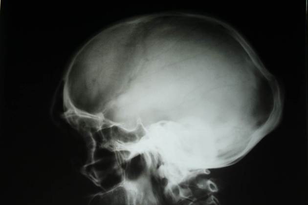
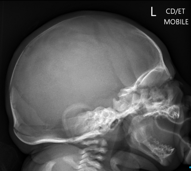
Bathrocephaly, also known as bathrocephalic occiputs, is a normal variation in skull shape, caused by an outward convex bulge of mid-portion of the occipital bone, often associated with a modification of the mendosal suture.
On this page:
Epidemiology
The true incidence of this disorder is unknown 1.
Rarely, there is an association with Hajdu-Cheney syndrome, which is characterized by hirsutism, joint laxity, acro-osteolysis, vertebral anomalies, bathrocephaly, and normal intelligence 4.
Clinical presentation
Bathrocephaly is of no clinical significance 2 and typically resolves with skull remodeling 2. Occasionally it may persist in adults 3.
Pathology
Bathrocephalic occiputs is a normal variation in skull shape, in which there is an outward convex bulge of mid-portion of the occipital bone 5. Bathrocephaly extends from the lambdoid to the mendosal suture. There is often a significant bulge of the interparietal portion of the occipital bone 3,6.
Etiology
The etiology of the condition is uncertain. Batrocephaly probably results from intrauterine modelling, with spontaneous remodeling being the rule 2,6. It can occur in babies who are a breech presentation in utero 2.
This disorder may be the result of incomplete fusion of the mendosal suture, which leads to bulging of the interparietal portion of the occipital bone. The skull protrusion slowly becomes less prominent and usually disappears 6. When it persists, it is associated with the characteristic head shape called bathrocephaly.
Radiographic features
Plain radiograph
Radiographs show a step-like bony protrusion of the posterior skull, with prominent occipital shelf, which should not be confused with pathologic changes. It is not associated with craniosynostosis 2.
CT
CT usually shows protrusion of the occipital bone between both parietal bones, with a step-like appearance of the occiput 6.
Treatment and prognosis
There is no evidence that intervention or surgical management of the disorder is necessary 1.
Differential diagnosis
cephalohematoma: ossified and non ossified
References
- 1. Justin Davanzo, Thomas Samson, R. Shane Tubbs, Elias Rizk. Bathrocephaly: a case report of a head shape associated with a persistent mendosal suture. (2019) Italian Journal of Anatomy and Embryology. 119 (3): 263-267. doi:10.13128/IJAE-15559
- 2. Kenneth F. Swaiman, Stephen Ashwal. Swaiman's Pediatric Neurology. (2012) ISBN: 9781437704358 - Google Books
- 3. Swischuk LE. Diagnóstico por imagens em neonatologia e pediatria. 3th ed. Rio de Janeiro, RJ: Livraria e editor Revinter. 1991: 851
- 4. Palav S, Vernekar J, Pereira S, Desai A. Hajdu-Cheney syndrome: a case report with review of literature. (2014) Journal of radiology case reports. 8 (9): 1-8. doi:10.3941/jrcr.v8i9.1833 - Pubmed
- 5. Dr. Emily R. Gallagher, Dr. Kelly N. Evans, Dr. Anne V. Hing, Dr. Michael L. Cunningham. Bathrocephaly: A Head Shape Associated with a Persistent Mendosal Suture:. (2013) The Cleft Palate-Craniofacial Journal. 50 (1): 104-8. doi:10.1597/11-153 - Pubmed
- 6. Alexander M. McKinney. Atlas of Normal Imaging Variations of the Brain, Skull, and Craniocervical Vasculature. (2017) ISBN: 9783319397900 - Google Books
- 7. Keats TE. An Atlas of Normal Roentgen Variants That May Simulate Disease. 2º ed. Chicago, IL: Year Book Medical Publishers, Inc. 1980: 52.
- 8. Caffey J. Pediatric X Ray Diagnosis. 6th ed. Chicago, IL: Year Book Medical Publishers, Inc. 1973: 14. (Alexander M. McKinney. Atlas of Normal Imaging Variations of the Brain, Skull, and Craniocervical Vasculature.
Incoming Links
Related articles: Anatomy: Head and neck
- skeleton of the head and neck
-
cranial vault
- scalp (mnemonic)
- fontanelle
-
sutures
- calvarial
- facial
- frontozygomatic suture
- frontomaxillary suture
- frontolacrimal suture
- frontonasal suture
- temporozygomatic suture
- zygomaticomaxillary suture
- parietotemporal suture (parietomastoid suture)
- occipitotemporal suture (occipitomastoid suture)
- sphenofrontal suture
- sphenozygomatic suture
- spheno-occipital suture (not a true suture)
- lacrimomaxillary suture
- nasomaxillary suture
- internasal suture
- basal/internal
- skull landmarks
- frontal bone
- temporal bone
- parietal bone
- occipital bone
- skull base (foramina)
-
facial bones
- midline single bones
- paired bilateral bones
- cervical spine
- hyoid bone
- laryngeal cartilages
-
cranial vault
- muscles of the head and neck
- muscles of the tongue (mnemonic)
- muscles of mastication
-
facial muscles
- epicranius muscle
- circumorbital and palpebral muscles
- nasal muscles
-
buccolabial muscles
- elevators, retractors and evertors of the upper lip
- levator labii superioris alaeque nasalis muscle
- levator labii superioris muscle
- zygomaticus major muscle
- zygomaticus minor muscle
- levator anguli oris muscle
- malaris muscle
- risorius muscle
- depressors, retractors and evertors of the lower lip
- depressor labii inferioris muscle
- depressor anguli oris muscle
- mentalis muscle
- compound sphincter
-
orbicularis oris muscle
- incisivus labii superioris muscle
- incisivus labii inferioris muscle
-
orbicularis oris muscle
- muscle of mastication
- modiolus
- elevators, retractors and evertors of the upper lip
- muscles of the middle ear
- orbital muscles
- muscles of the soft palate
- pharyngeal muscles
- suprahyoid muscles
- infrahyoid muscles
- intrinsic muscles of the larynx
- muscles of the neck
- platysma muscle
- longus colli muscle
- longus capitis muscle
- scalenus anterior muscle
- scalenus medius muscle
- scalenus posterior muscle
- scalenus pleuralis muscle
- sternocleidomastoid muscle
-
suboccipital muscles
- rectus capitis posterior major muscle
- rectus capitis posterior minor muscle
- obliquus capitis superior muscle
- obliquus capitis inferior muscle
- accessory muscles of the neck
- deep cervical fascia
-
deep spaces of the neck
- anterior cervical space
- buccal space
- carotid space
- danger space
- deep cervical fascia
- infratemporal fossa
- masticator space
- parapharyngeal space
- stylomandibular tunnel
- parotid space
- pharyngeal (superficial) mucosal space
- perivertebral space
- posterior cervical space
- pterygopalatine fossa
- retropharyngeal space
- suprasternal space (of Burns)
- visceral space
- surgical triangles of the neck
- orbit
- ear
- paranasal sinuses
- upper respiratory tract
- viscera of the neck
- blood supply of the head and neck
-
arterial supply
-
common carotid artery
- carotid body
- carotid bifurcation
- subclavian artery
- variants
-
common carotid artery
- venous drainage
-
arterial supply
- innervation of the head and neck
-
cranial nerves
- olfactory nerve (CN I)
- optic nerve (CN II)
- oculomotor nerve (CN III)
- trochlear nerve (CN IV)
-
trigeminal nerve (CN V) (mnemonic)
- trigeminal ganglion
- ophthalmic division
- maxillary division
- mandibular division
- abducens nerve (CN VI)
- facial nerve (CN VII)
-
vestibulocochlear nerve (CN VIII)
- vestibular ganglion (Scarpa's ganglion)
- glossopharyngeal nerve (CN IX)
- vagus nerve (CN X)
- (spinal) accessory nerve (CN XI)
- hypoglossal nerve (CN XII)
- parasympathetic ganglia of the head and neck
- cervical sympathetic ganglia
- greater occipital nerve
- third occipital nerve
-
cervical plexus
- muscular branches
- longus capitis
- longus colli
- scalenes
- geniohyoid
- thyrohyoid
-
ansa cervicalis
- omohyoid (superior and inferior bellies separately)
- sternothyroid
- sternohyoid
- phrenic nerve
- contribution to the accessory nerve (CN XI)
- cutaneous branches
- muscular branches
- brachial plexus
- pharyngeal plexus
-
cranial nerves
- lymphatic drainage of the head and neck
- embryological development of the head and neck


 Unable to process the form. Check for errors and try again.