
The branchial (or pharyngeal) apparatus is the complex region in the developing embryo between the head and chest that develops in the fourth week and provides bilateral ridges and valleys that subsequently develop into numerous anatomic structures of the head, face, palate and anterior neck. The development of structures from the apparatus helps explain the complex cranial nerve distribution in some regions.
The apparatus resembles the branchia (gills) of fish and amphibians, which obviously don't form in the human. Hence the description of pharyngeal arches etc. is more correct and advised.
Structure
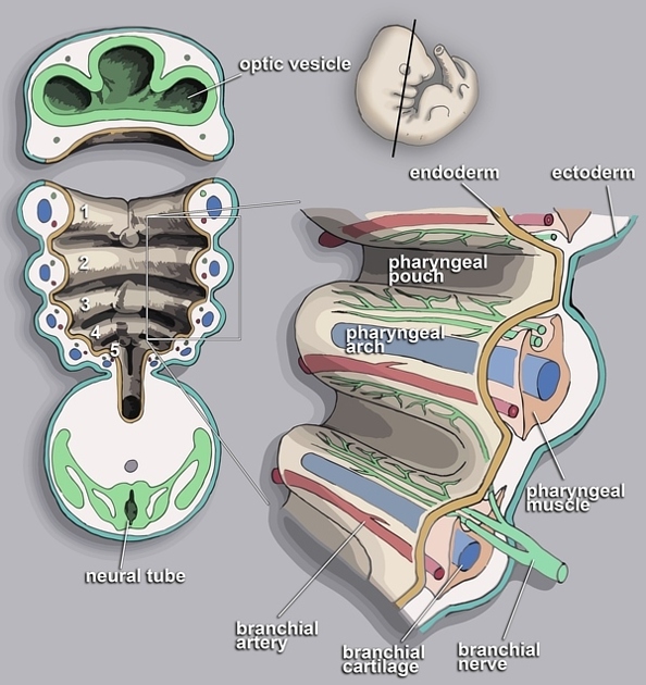
The apparatus consists of paired symmetrical pharyngeal arches, pouches, grooves and membranes that develop from the ectoderm and in part from the pharyngeal part of the primitive foregut (see figure 1). They start forming in week 4 and by about week 7 the differentiation and migration of structures from the branchial apparatus is complete.
-
pharyngeal arches are transverse swellings that laterally border the proximal foregut, each is separated from adjacent arches by the branchial clefts
contains both mesoderm (forming muscles and vessels) and mesenchyme (neural crest cells which form bones)
each arch has a cranial nerve associated with it, along with a cartilage piece and artery associated with aortic arch development, some of which obliterate
the fifth arch does not develop but for comparative anatomy reasons, the sixth arch retains its name
-
pharyngeal pouches form as endodermal invaginations from the lateral wall of the proximal primitive foregut
these penetrate the mesenchyme but do not form an open communication with the external clefts
pharyngeal or branchial clefts (or grooves) form as ectodermal invaginations on the external surface of the embryo, between the arches
pharyngeal membranes are formed between the pharyngeal pouch and groove
Pharyngeal arches
-
the first pharyngeal arch is the largest and forms a dorsal maxillary process and a ventral mandibular process, which contains Meckel's cartilage. It contributes to the development of the face and several facial bones as well as the temporal bone
cranial nerve: CN Vc
-
mesodermal derivatives: tensor tympani, muscles of mastication, mylohyoid, anterior belly of digastric, tensor veli palatini
artery: first aortic arch (temporary)
neural crest cell derivatives: incus, malleus, anterior ligament of malleus, sphenoid spine, sphenomandibular ligament and the genial tubercle of the mandible
-
the second pharyngeal arch contains Reichert's cartilage
cranial nerve: CN VII
-
mesodermal derivatives: stapedius, stylohyoid, muscles of facial expression, posterior belly of digastric
artery: stapedial artery (temporary)
neural crest cell derivatives: stapes, styloid process of the temporal bone, stylohyoid ligament, lesser horn and upper part of the body of the hyoid bone
-
the third pharyngeal arch is small
cranial nerve: CN IX
-
mesodermal derivatives: stylopharyngeus
neural crest cell derivatives: greater horn and lower part of body of hyoid bone
-
the fourth pharyngeal arch cartilage fuses with that of the sixth arch
cranial nerve: CN X (pharyngeal branch and superior laryngeal nerve)
-
mesodermal derivatives: pharyngeal constrictor muscles, extrinsic laryngeal muscles, levator veli palatini
artery: on the right the first part of the subclavian artery, on the left the aortic arch (between the left CCA and left subclavian artery)
neural crest cell derivatives: thyroid, corniculate and cuneiform cartilages
the fifth pharyngeal arch does not exist in humans
-
the sixth pharyngeal arch
cranial nerve: CN X (recurrent laryngeal nerve)
-
mesodermal derivatives: intrinsic laryngeal muscles
artery: ductus arteriosus
neural crest cell derivatives: arytenoid cartilages
Pharyngeal pouches
the first pharyngeal pouch forms as a diverticulum called the tubotympanic recess and gives rise to the epithelium of the auditory tube and middle ear cavity
the second pharyngeal pouch gives rise to the epithelium of the palatine tonsil
the third pharyngeal pouch has ventral and dorsal wings and gives rise to the thymus (from the ventral wing) and inferior parathyroid gland (from the dorsal wing)
the fourth pharyngeal pouch gives rise to the superior parathyroid gland
the fifth pharyngeal pouch is atypical and often considered to be part of the fourth pouch; it gives rise to the ultimobranchial body which contributes to the parafollicular (C) cells of the thyroid gland responsible for the secretion of calcitonin
Pharyngeal cleft
the first pharyngeal cleft forms the epithelium of the external auditory meatus and the external epithelium of the tympanic membrane
the second to fourth pharyngeal clefts fuse and form the cervical sinus (of His), which typically obliterates
Pharyngeal membranes
the first pharyngeal membrane forms the tympanic membrane
the second to fifth pharyngeal membranes are obliterated
Related pathology
-
first branchial cleft anomalies
-
first branchial arch anomalies
-
second branchial cleft anomalies
-
third branchial cleft anomalies
-
third branchial arch anomalies
-
third branchial pouch anomalies
ectopic inferior parathyroid glands
ectopic thymus
-
fourth branchial cleft anomalies
-
fourth branchial pouch anomalies
ectopic superior parathyroid glands


 Unable to process the form. Check for errors and try again.