
Breast venous malformations (also known as breast hemangiomas) are benign vascular lesions occurring within breast tissue.
Most breast venous malformations are so called cavernous malformations, which are found throughout the body. For a general discussion please refer to the general article on cavernous venous malformation.
On this page:
Terminology
It is important to note that according to newer nomenclature (ISSVA classification of vascular anomalies) these lesions are merely known as slow flow venous malformations. Having said that it is probably helpful in reports to include the word 'hemangioma' as this term is ubiquitous in the literature and most familiar to many clinicians. The remainder of this article often uses the term 'breast hemangioma' for consistency with the majority of the existing literature.
Epidemiology
Breast hemangiomas are reported to be found in ~ 1.2% of mastectomy specimens and 11% of post-mortem specimens. They can occur at almost any age with an age range between 18 months and 82 years.
Pathology
The venous malformations that occur within the breast are most commonly the cavernous type 2. While lesions are most often superficial within the breast tissue, there is no recognized predilection towards any particular location within the breast. Histologically they are similar to cavernous venous malformations (hemangiomas) at other sites 9 and comprise of lobulated cluster of non-neoplastic vessels filled with slow moving blood.
Radiographic features
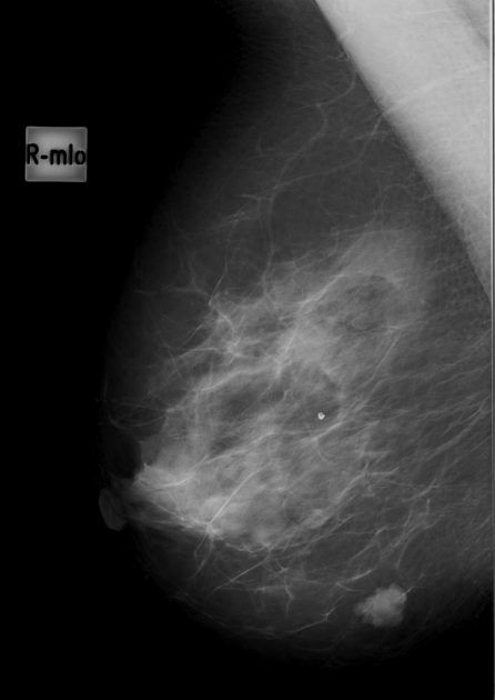
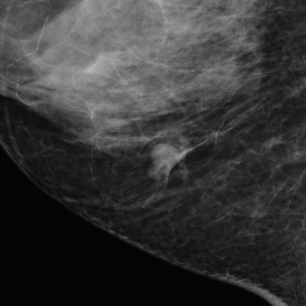
Mammography
Mammographic appearances are generally non-specific. Typical appearance is of oval or lobular isodense masses with well-circumscribed margins. Calcification (phleboliths) is variably present.
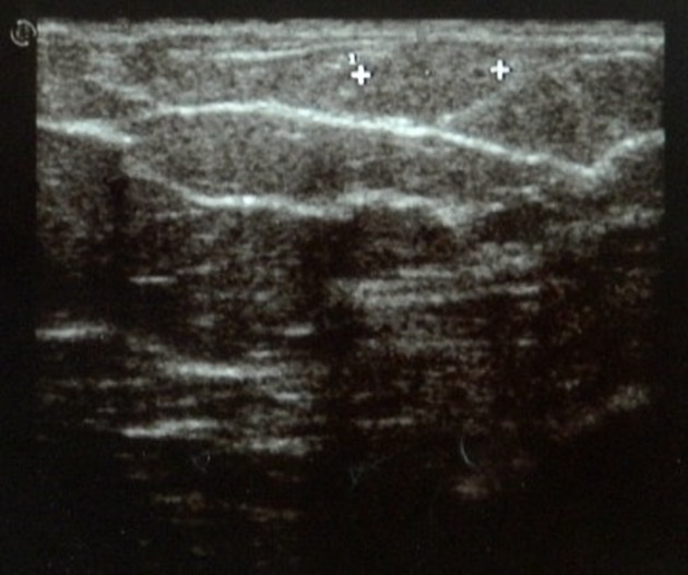
Breast ultrasound
A hemangioma may appear as a lobulated, superficial, well-circumscribed, solid mass that is predominantly hypoechoic. It may contain areas of calcification. Hyperechoic nodules also have been described.
Treatment and prognosis
Given the non specific imaging appearance, diagnosis is commonly made following a core biopsy.
Consequent management may be either conservative with radiologic followup, or with excision biopsy. Generally lesions with classic imaging and histopathologic features may be treated conservatively, whereas excision is recommended in radiologically or pathologically atypical cases, where angiosarcoma needs to be excluded10.
Progression of hemangioma to angiosarcoma is controversial. Given the frequency of occult hemangiomas on postmortem specimens, and the rarity of angiosarcomas, malignant transformation is extremely rare, if exists at all1.
Differential diagnosis
- fibroadenoma: may have overlapping imaging features
- complex cyst: may have overlapping imaging features
- angiosarcoma: diagnosis requiring exclusion in atypical cases10


 Unable to process the form. Check for errors and try again.