
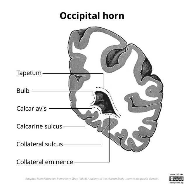
Calcar avis
Citation, DOI, disclosures and article data
At the time the article was created Pradeep J. Gamage had no recorded disclosures.
View Pradeep J. Gamage's current disclosuresAt the time the article was last revised Frank Gaillard had the following disclosures:
- Biogen Australia Pty Ltd, Investigator-Initiated Research Grant for CAD software in multiple sclerosis: finished Oct 2021 (past)
These were assessed during peer review and were determined to not be relevant to the changes that were made.
View Frank Gaillard's current disclosures- Hippocampus minor
Calcar avis is an elevation of white matter projecting from the medial wall of the occipital horn of the lateral ventricle. It is variably conspicuous, depending on how deep the calcarine sulcus is.
On this page:
Gross anatomy
The calcar avis is located on the medial wall of the occipital horn, near the junction with the trigone of the lateral ventricles. It is formed by white matter separating the ventricle from the calcarine sulcus and, thus, is more prominent if the sulcus is deep 1-3. It is located below the bulb of the occipital horn, formed by forceps major, and above the collateral eminence, formed by white matter separating the ventricle from the collateral sulcus 2.
Radiographic features
Ultrasound
In a coronal plane of the neonatal brain, the calcar avis can mimic an intraventricular hemorrhage. Continuation of it as an echogenic fissure as the calcarine fissure and normal vascularity aids in differentiating the calcar avis from a blood clot 1.
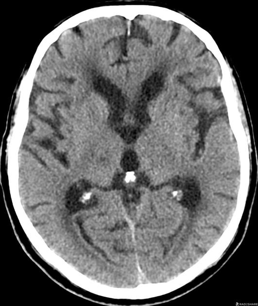
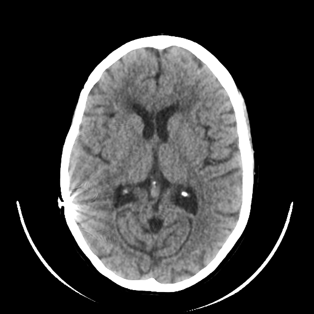
CT
On axial reconstructions, the calcar avis may mimic intraventricular hemorrhage in the occipital horns. This structure can also mimic grey matter heterotopia and schizencephaly ref.
History and etymology
The calcar avis was previously known as the hippocampus minor (in contrast to hippocampus major, now simply the hippocampus) 1.
References
- 1. Owen C, Howard A, Binder D. Hippocampus Minor, Calcar Avis, and the Huxley-Owen Debate. Neurosurgery. 2009;65(6):1098-105. doi:10.1227/01.neu.0000359535.84445.0b - Pubmed
- 2. Flores L. Occipital Lobe Morphological Anatomy: Anatomical and Surgical Aspects. Arq Neuro-Psiquiatr. 2002;60(3A):566-71. doi:10.1590/s0004-282x2002000400010
- 3. Savas R & Sener R. Deep Calcarine Sulcus and Prominent Calcar Avis. J Neuroradiol. 1998;25(2):144-6. - Pubmed
Incoming Links
Related articles: Anatomy: Brain
-
brain
- grey matter
- white matter
-
cerebrum
-
cerebral hemisphere (telencephalon)
- cerebral lobes and gyri
- frontal lobe
- parietal lobe
-
occipital lobe
- occipital pole
- lingual gyrus
- fusiform gyrus (Brodmann area 37)
- calcarine (visual) cortex
- cuneus
- temporal lobe
- basal forebrain
- limbic system
- insula
-
cerebral sulci and fissures (A-Z)
- calcarine fissure
- callosal sulcus
- central (Rolandic) sulcus
- cingulate sulcus
- collateral sulcus
- inferior frontal sulcus
- inferior occipital sulcus
- inferior temporal sulcus
- interhemispheric fissure
- intraparietal sulcus
- lateral (Sylvian) sulcus
- lateral occipital sulcus
- marginal sulcus
- occipitotemporal sulcus
- olfactory sulcus
- paracentral sulcus
- paraolfactory sulcus
- parieto-occipital fissure
- posterior parolfactory sulcus
- precentral sulcus
- preoccipital notch
- postcentral sulcus
- rhinal sulcus
- rostral sulcus
- subparietal sulcus
- superior frontal sulcus
- superior occipital sulcus
- superior temporal sulcus
- cortical histology
- cerebral lobes and gyri
- white matter tracts
- deep grey matter
-
pituitary gland
- posterior pituitary and stalk (part of diencephalon)
- anterior pituitary
- inferior hypophyseal arterial circle
- diencephalon
-
cerebral hemisphere (telencephalon)
-
brainstem
- midbrain (mesencephalon)
- pons (part of metencephalon)
- medulla oblongata (myelencephalon)
- white matter
-
grey matter
- non-cranial nerve
-
cranial nerve nuclei
- oculomotor nucleus
- Edinger-Westphal nucleus
- trochlear nucleus
- motor nucleus of CN V
- mesencephalic nucleus of CN V
- main sensory nucleus of CN V
- spinal nucleus of CN V
- abducent nucleus
- facial nucleus
- superior salivatory nucleus
- cochlear nuclei
- vestibular nuclei
- inferior salivatory nucleus
- solitary tract nucleus
- ambiguus nucleus
- dorsal vagal motor nucleus
- hypoglossal nucleus
-
cerebellum (part of metencephalon)
- vermis
- cerebellar hemisphere
- cerebellar peduncles
- cranial meninges (meninx primitiva)
- CSF spaces
-
cranial nerves (mnemonic)
- olfactory nerve (CN I)
- optic nerve (CN II)
- oculomotor nerve (CN III)
- trochlear nerve (CN IV)
- trigeminal nerve (CN V) (mnemonic)
- abducens nerve (CN VI)
- facial nerve (CN VII) (segments mnemonic | branches mnemonic)
-
vestibulocochlear nerve (CN VIII)
- vestibular ganglion (Scarpa's ganglion)
- glossopharyngeal nerve (CN IX)
- vagus nerve (CN X)
- spinal accessory nerve (CN XI)
- hypoglossal nerve (CN XII)
- functional neuroanatomy
- CNS development
- cerebral vascular supply
- arteries
- vascular territories
-
circle of Willis
- internal carotid artery (ICA) (segments)
- vertebral artery
-
normal variants
- intracranial arterial fenestration
- internal carotid artery (ICA)
- anterior cerebral artery (ACA)
- middle cerebral artery (MCA)
- posterior cerebral artery (PCA)
- basilar artery
- persistent carotid-vertebrobasilar artery anastomoses (mnemonic)
- vertebral artery
- ophthalmic artery
-
cerebral venous system
-
dural venous sinuses
- basilar venous plexus
- cavernous sinus (mnemonic)
- clival diploic veins
- inferior petro-occipital vein
- inferior petrosal sinus
- inferior sagittal sinus
- intercavernous sinus
- internal carotid artery venous plexus of Rektorzik
- jugular bulb
- marginal sinus
- occipital sinus
- sigmoid sinus
- sphenoparietal sinus
- straight sinus
- superior petrosal sinus
- superior sagittal sinus
- torcula herophili
- transverse sinus
-
cerebral veins
-
superficial veins of the brain
- superior cerebral veins (superficial cerebral veins)
- inferior cerebral veins
- superficial middle cerebral vein
- superior anastomotic vein (of Trolard)
- inferior anastomotic vein (of Labbe)
-
superficial veins of the brain
-
deep veins of the brain
- great cerebral vein (of Galen)
- venous circle of Trolard
- normal variants
-
dural venous sinuses
- arteries
- glymphatic pathway


 Unable to process the form. Check for errors and try again.