
Chronic venous insufficiency occurs due to inadequate functioning of venous walls and/or valves in the lower limbs resulting in excessive pooling of blood.
On this page:
Clinical presentation
Symptoms of chronic venous insufficiency include heaviness, tension, swelling feeling, aching, itching and/or cramps 7.
Pathology
The condition results from venous hypertension which in turn is usually caused by reflux in the superficial venous compartment. Less common causes include:
deep venous compression
post-thrombotic stenosis or occlusion
deep venous reflux
venous hypertension caused by vascular malformations, arteriovenous fistulae, and neuromuscular disorders (rare)
Classification
Radiographic features
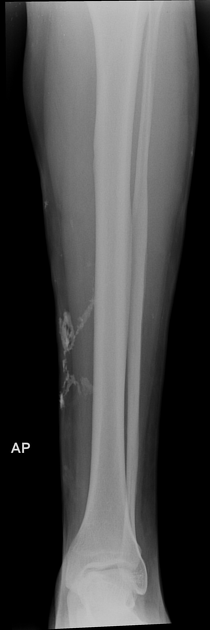
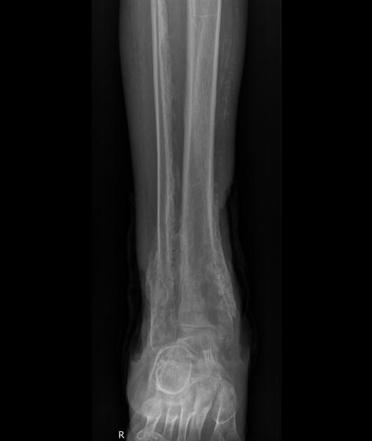
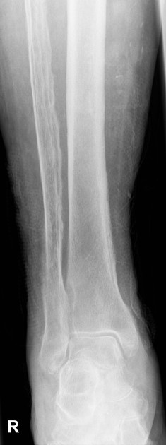
Plain radiograph
Findings are non-specific but most commonly are seen in the leg 5,6:
solid undulating periosteal reaction, often symmetrical
dystrophic soft tissue calcification
soft tissue swelling from subcutaneous edema
Ultrasound
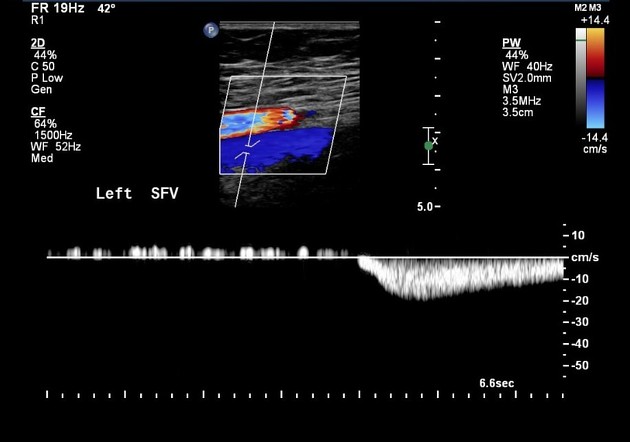
Venous Doppler ultrasound
Considered the primary imaging modality of choice. Typically the great saphenous vein and the small saphenous vein and their primary tributaries are assessed.
The presence of reflux is determined by the direction of flow because any significant flow toward the feet is suggestive of reflux. The duration of reflux is known as the "reflux time" (replacing the commonly used "valve closure time"):
reflux time of >0.5 seconds for superficial veins and 1.0 second for deep veins has been used to diagnose the presence of reflux 8, although a more refined definition with a variable “cut off” based on location has been suggested ref
the longer the duration of reflux or the greater the reflux time implies more severe disease
Venous duplex imaging may provide information about local valve function to construct an anatomic map of disease in terms of the systems and levels of involvement.
The presence and location of perforators are also documented. The patient should be able to stand for this procedure.
Complications
venous ulceration in ~20% 8


 Unable to process the form. Check for errors and try again.