
Craniosynostosis (plural: craniosynostoses) refers to the premature closure of the cranial sutures. The skull shape then undergoes characteristic changes depending on which suture(s) close early.
On this page:
Epidemiology
There is a 3:1 male predominance with an overall incidence of 1 in 2000-2500. 8% of cases are syndromic or familial.
Associations
Most occur as isolated anomalies but syndromic associations can be seen in a small proportion of cases (~10%):
Pathology
Aetiology
Primary forms are either sporadic or familial. Secondary craniosynostosis occurs in relation to a variety of causes:
-
endocrine disorders
hypophosphataemia
hypercalcaemia
-
haematologic disorders causing bone marrow hyperplasia
-
inadequate brain growth
shunted hydrocephalus
Ages of normal sutural/fontanelle closure
metopic: 3-9 months
anterior fontanelle: 18-24 months
sphenosquamosal: 6-10 years
sphenofrontal: approximately 15 years
occipitomastoid: approximately 16 years
sagittal: approximately 22 years
coronal: approximately 24 years
lambdoid: approximately 26 years
squamosal: approximately 60 years
Types
brachycephaly: bicoronal and/or bilambdoid sutures
scaphocephaly/dolichocephaly: sagittal suture
-
plagiocephaly: unilateral coronal and lambdoid sutures
frontal plagiocephaly: unilateral coronal suture
occipital plagiocephaly: unilateral lambdoid suture
pachycephaly: lambdoid suture
oxycephaly/turricephaly: sagittal, coronal and lambdoid sutures (tower like skull)
cloverleaf skull/Kleeblattschädel: intrauterine sagittal, coronal, lambdoid sutures (most severe)
harlequin eye: ipsilateral coronal suture
progressive postnatal pansynostosis: a rare form of craniosynostosis which involves late (postnatal) fusion of all cranial sutures 9
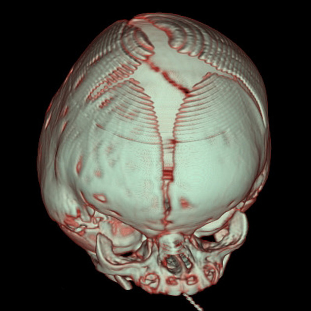
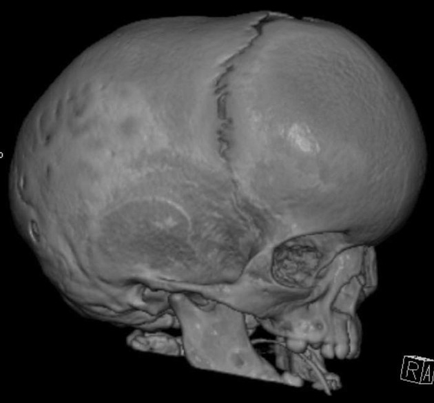
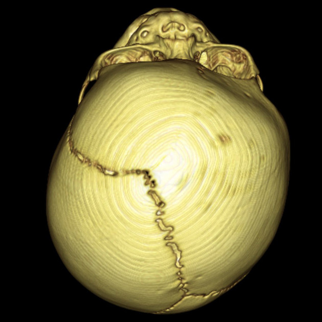
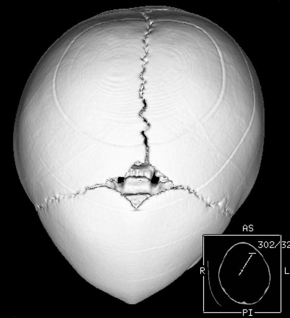
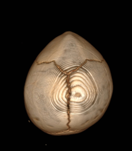
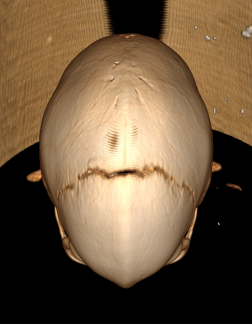
The sagittal suture is most commonly involved (≈50%), where the lateral growth of the skull is arrested while anteroposterior growth continues, producing a narrowly elongated skull known as scaphocephaly (meaning boat-shaped) or dolichocephaly (from the ancient Greek for long, δολιχός: dolichos).
The next most common sutures in terms of involvement are:
coronal (~20%)
lambdoid (~5%)
metopic (~5%)
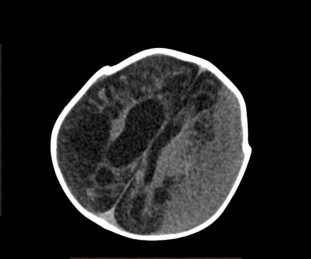
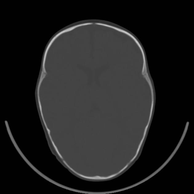
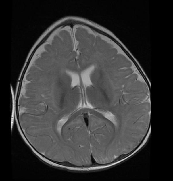
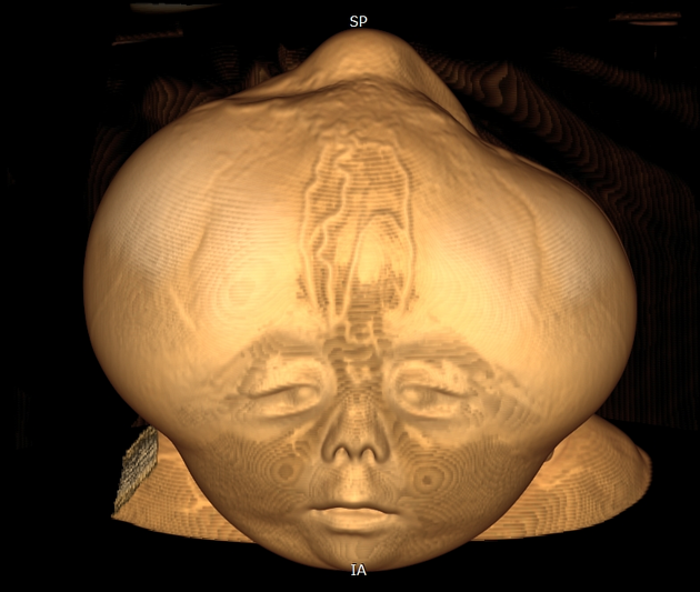
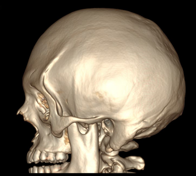
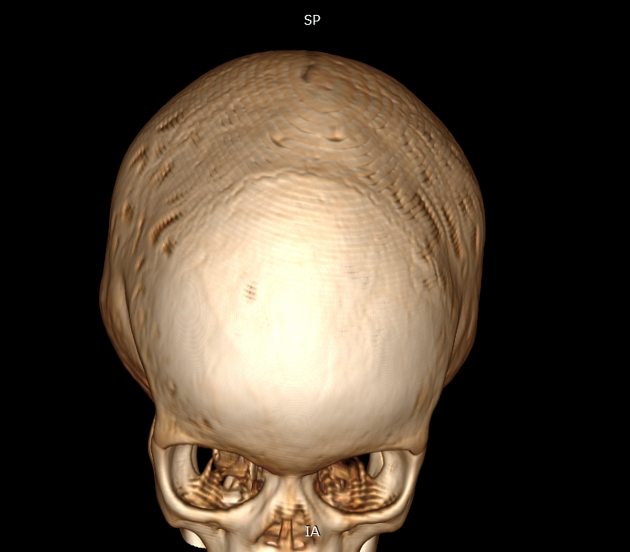
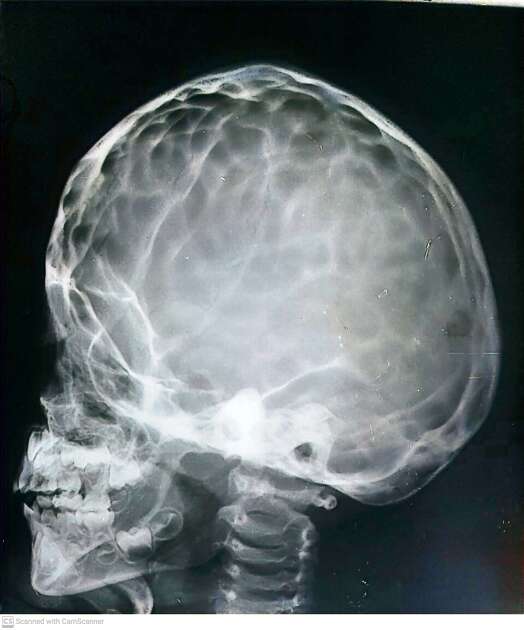
Radiographic features
Restriction of skull growth is perpendicular to the affected suture line. Characteristic dysmorphic head shapes are associated with each type of craniosynostosis.
Ultrasound
Ultrasound can be used as a screening tool or in clinically-subtle cases and can reduce radiation exposure in infants to cases with inconclusive findings 11.
General features include:
-
sutures are normally hypoechoic
there may be a loss of normally decreased echogenicity in the region of the fusion
lack of suture patency
ridging of the sutures
CT
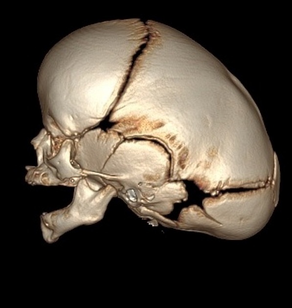
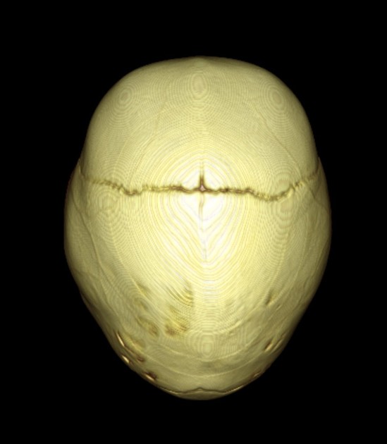
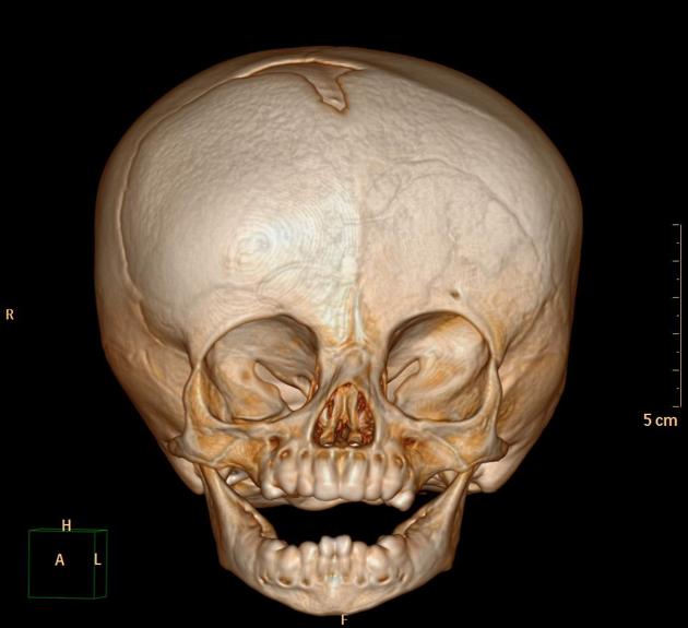
Low-dose CT with 3D image reformations is the best modality for the evaluation of skull sutures 5.
Treatment and prognosis
Treatment is often with a cranioplasty. Abnormal intracranial pressure may affect neurocognition.
History and etymology
Craniosynostosis was first accurately characterised by Rudolph Virchow in 1851 10.


 Unable to process the form. Check for errors and try again.