
Diagnostic HRCT criteria for usual interstitial pneumonia (UIP) pattern - ATS/ERS/JRS/ALAT (2018)
Updates to Article Attributes
SpecificAs a part of international evidence based guidelines adopted by collaborative effort of American Thoracic Society, the European Respiratory Society, the Japanese Respiratory Society and the Latin American Thoracic association, specific diagnostic HRCT criteria for usual interstitial pneumonia (UIP) pattern have beenwere adopted by international consensusin 2011. At the time of ATS/ERS/JRS/ALAT to categorise CT-based certainty forwriting (mid 2016), this is the UPmost widely accepted classification system.
Classification
-
UIP pattern
into three levels:definite(definite) -
possible UIP
,probable -
inconsistent UIP
andinconsistent with UIP.pattern
This helps radiologists to determine the certainty of UIP diagnosis based on HRCT chest findings. The importance of this guideline is that definite UIP pattern on chest HRCT precludes the need for tissue diagnosis 1,2. However unfortunately up to 20 % of inconsistent with UIP group (or actually atypical UIP) can be UIP on biopsy or IPFidiopathic pulmonary fibrosis (IPF) in clinical course.
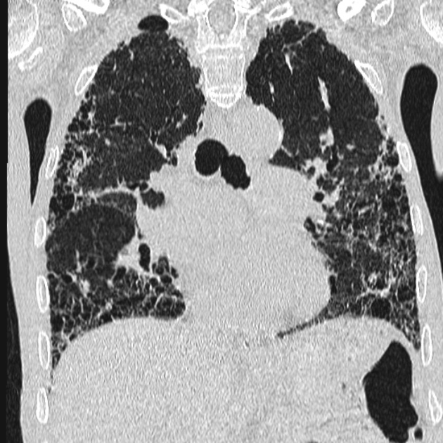
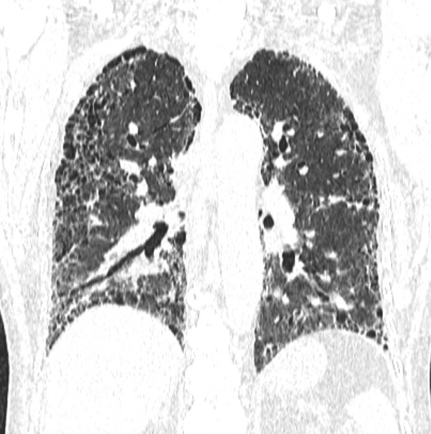
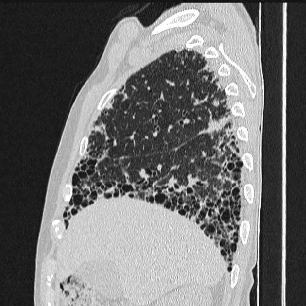
UIP pattern
-
Allall four features present:- subpleural, basal predominance
- reticular abnormality
- honeycombing +/- traction bronchiectasis
- absence of features listed as
"inconsistent'inconsistent with UIP"' (see below)
Possible UIP pattern
-
Allall three features present*- subpleural, basal predominance
- reticular abnormality
- absence of features listed as
"inconsistent'inconsistent with UIP"' (see below)
* honeycombing is absent
Inconsistent with UIP pattern
-
Anyany one of the following seven features present:- upper or mid-lung predominance
- peribronchovascular predominance
- extensive ground glass abnormality (i.e. more than reticular abnormality)
- profuse micronodules (bilateral, predominantly upper lobes)
- discrete cysts (multiple, bilateral, away from honeycombing)
- diffuse mosaic attenuation / air-trapping (bilateral in ≥3 lobes)
- consolidation in bronchopulmonary segment(s) or lobe(s)


 Unable to process the form. Check for errors and try again.