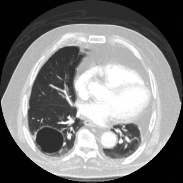
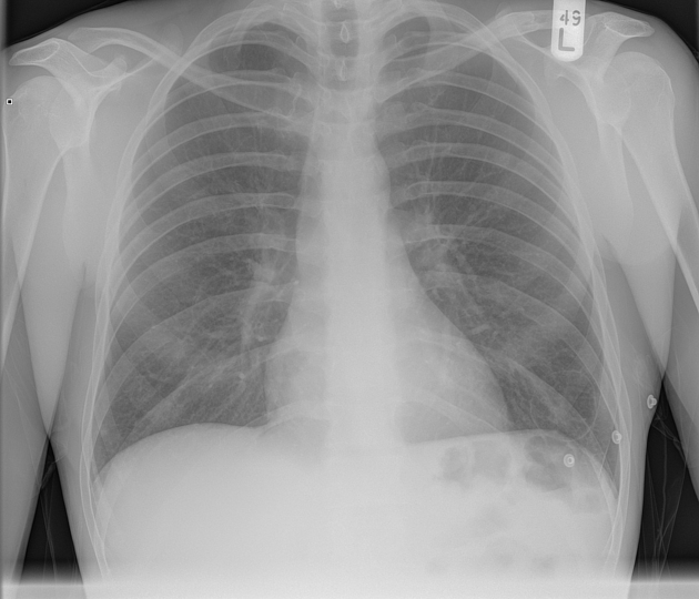
A pulmonary cyst is any well-circumscribed gas-containing structure within lung parenchyma with a thin, typically regular wall. Occasionally a cyst may contain fluid or solid material instead of gas 10.
On this page:
Terminology
The term ‘cystic’ denotes lesions with central gas attenuation contained by a wall, regardless of etiology. The wall thickness and regularity may vary. The morphology of cystic lesions such as blebs, bullae and cysts overlaps and there are no objective criteria to quantify wall thickness and no consensus regarding definitions 10.
Epidemiology
A few cysts may develop with aging and are generally seen in patients >40 years of age with a low BMI 9.
Pathology
Pulmonary cysts can be congenital or acquired and can be caused by:
check-valve obstruction with distal airspace dilatation
bronchial wall necrosis
protease-mediated parenchymal destruction 10
Multiple lung cysts in a child may be associated with an underlying process although this is rare, e.g. pleuropulmonary blastomas 1.
Differential diagnosis
Other thin-walled cystic spaces in the lungs 6,7:
bleb: pleural/subpleural, usually <1 cm diameter
bulla: pleural/subpleural
honeycombing: subpleural stacks of cysts, typically 3-10 mm diameter with walls 1-3 mm in thickness
pneumatocele: usually transient cystic airspace within the lung, usually due to pneumonia or trauma
Pulmonary cysts have many minics:
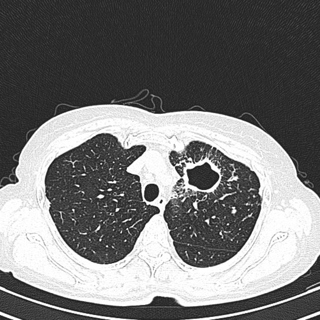
pulmonary cavity: surrounded by mass, nodule, or consolidation, creating wall thickness >2-4 mm 4-6
emphysema: lucencies without wall and with central vessel
cystic bronchiectasis: contiguous with other airways
occasionally lung cancer may appear as thin walled cystic lung cancer


 Unable to process the form. Check for errors and try again.