
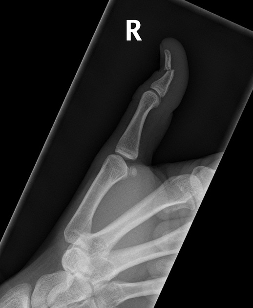
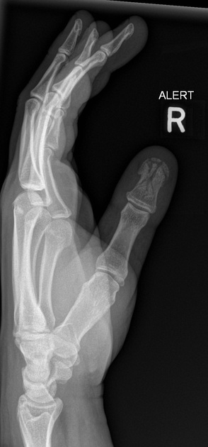
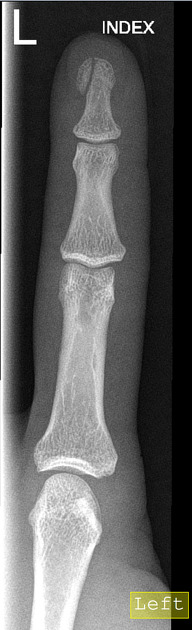
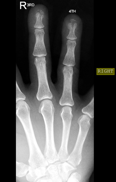
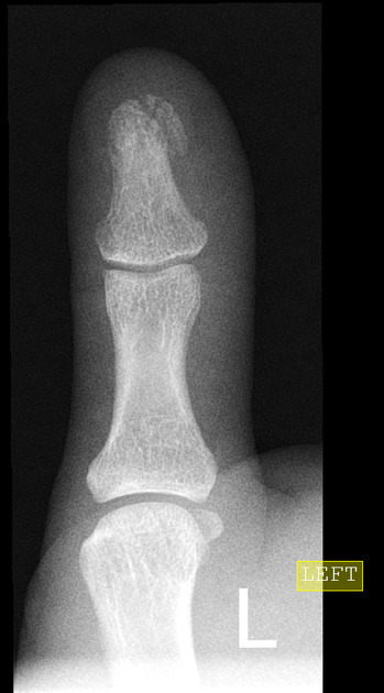
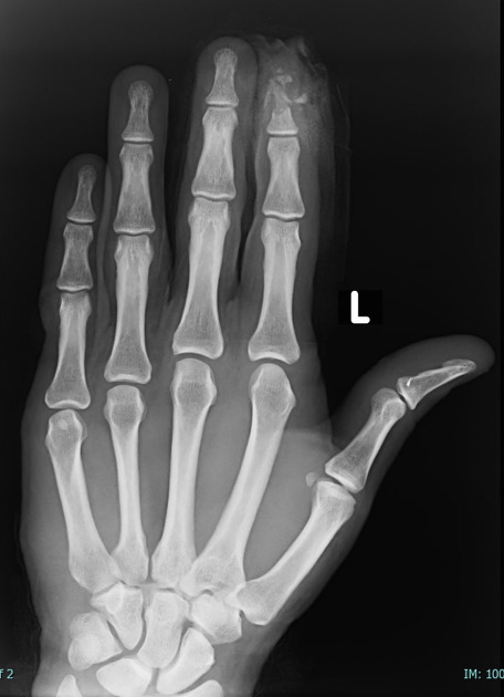
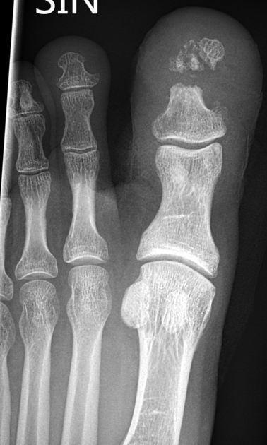
Distal phalanx fracture
Citation, DOI, disclosures and article data
At the time the article was created Tom O'Graphy had no recorded disclosures.
View Tom O'Graphy's current disclosuresAt the time the article was last revised Mostafa Elfeky had no financial relationships to ineligible companies to disclose.
View Mostafa Elfeky's current disclosures- Phalangeal tuft fracture
- Distal phalangeal fracture
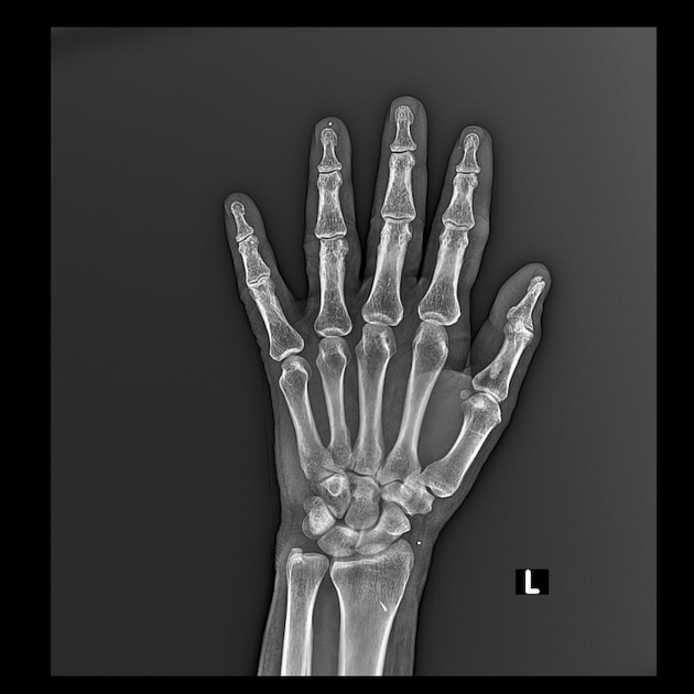
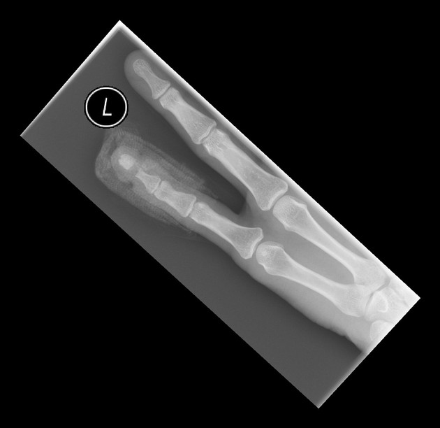
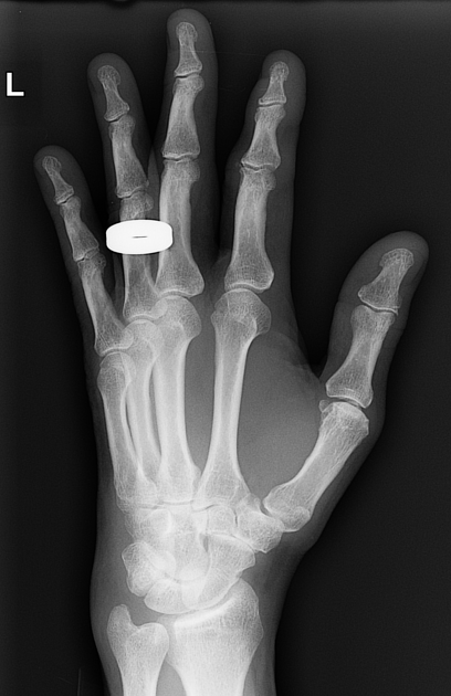
Distal phalanx fractures are among the most common fractures in the hand.
They represent > 50% of all phalangeal fractures and frequently involve the ungual tuft 1.
They are frequently related to sports, with lesions such as the mallet finger and the Jersey finger. When associated with a crush injury, open fracture is more likely.
Radiographic features
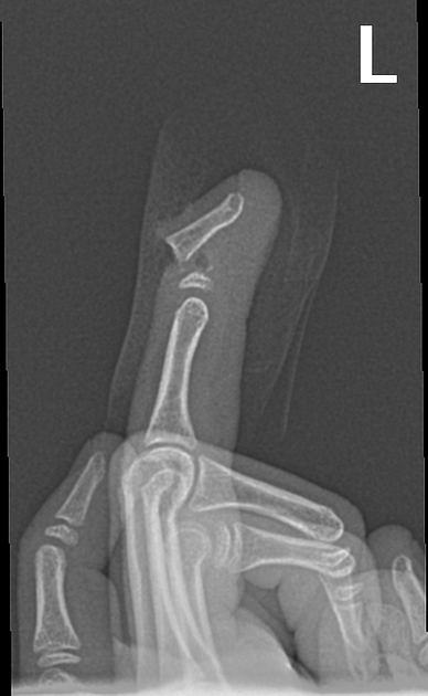
Plain radiographs form the mainstay of imaging distal phalanx fractures. The presence or absence of an intra-articular component, degree of comminution, and fracture displacement should be assessed when formulating a report.
Fracture angulation can be difficult to assess radiologically and is best assessed on clinical examination.
Treatment and prognosis
The majority of distal phalanx fractures are minimally displaced and may be treated conservatively. Closed fractures are generally stable, especially when they do not involve the articular surface. A mallet splint is often used in these cases.
Fractures at the base of the distal phalanx are often unstable due to the fact these are the insertions sites for both the flexor and extensor tendon, however splinting of these fractures, granted they are closed has favourable outcomes 3.
As in all cases of trauma, the importance of recognition of open distal phalanx fractures is due to the increased risk of contamination and, hence, infection. Extensive wound irrigation, antibiotic cover, and tetanus booster prophylaxis must be considered to mitigate this risk. Surgery for nailbed repair and/or Kirschner wire fixation will be required in more complex cases.
Extensively comminuted fractures and/or fractures with soft tissue injury to the nail bed should be considered as an open fracture and clinically treated as such (with antibiotics). Failure to do so may result in distal phalanx osteomyelitis, and additional complications such as growth arrest.
Quiz questions
References
- 1. Yoong P, Goodwin RW, Chojnowski A. Phalangeal fractures of the hand. (2010) Clinical radiology. 65 (10): 773-80. doi:10.1016/j.crad.2010.04.008 - Pubmed
- 2. Peterson JJ, Bancroft LW. Injuries of the fingers and thumb in the athlete. (2006) Clinics in sports medicine. 25 (3): 527-42, vii-viii. doi:10.1016/j.csm.2006.02.001 - Pubmed
- 3. Carpenter S, Rohde RS. Treatment of phalangeal fractures. (2013) Hand clinics. 29 (4): 519-34. doi:10.1016/j.hcl.2013.08.006 - Pubmed
Incoming Links
- Mallet finger
- Comminuted fracture of the distal phalanx of great toe
- Distal phalanx fracture
- Distal phalangeal fracture
- Mallet finger
- Distal phalanx fracture
- Distal phalanx fracture
- Distal phalanx fracture
- Big toe fracture
- Mallet finger
- Open fracture of the distal phalanx
- Toe fracture
- Distal phalanx fracture
- Distal phalanx fracture
- Distal phalanx fracture
- Mallet finger
- Distal phalanx fracture
- Big toe comminuted fracture
- Mallet finger
- Distal phalanx fracture
Related articles: Fractures
-
fracture
- terminology
- fracture location
- diaphyseal fracture
- metaphyseal fracture
- physeal fracture
- epiphyseal fracture
- fracture types
- avulsion fracture
- articular surface injuries
- complete fracture
- incomplete fracture
- infraction
- compound fracture
- pathological fracture
- stress fracture
- fracture displacement
- fracture location
- fracture healing
- skull fractures
-
facial fractures
- fractures involving a single facial buttress
- alveolar process fractures
- frontal sinus fracture
- isolated zygomatic arch fractures
- mandibular fracture
- nasal bone fracture
- orbital blow-out fracture
- paranasal sinus fractures
- complex fractures
- dental fractures
- fractures involving a single facial buttress
-
spinal fractures
- classification (AO Spine classification systems)
-
cervical spine fracture classification systems
- AO classification of upper cervical injuries
- AO classification of subaxial injuries
- Anderson and D'Alonzo classification (odontoid fracture)
- Roy-Camille classification (odontoid process fracture)
- Gehweiler classifcation (atlas fractures)
- Levine and Edwards classification (hangman fracture)
- Allen and Ferguson classification (subaxial spine injuries)
- subaxial cervical spine injury classification (SLIC)
- thoracolumbar spinal fracture classification systems
- three column concept of spinal fractures (Denis classification)
- classification of sacral fractures
-
cervical spine fracture classification systems
- spinal fractures by region
- spinal fracture types
- classification (AO Spine classification systems)
- rib fractures
- sternal fractures
-
upper limb fractures
- classification
- Rockwood classification (acromioclavicular joint injury)
- AO classification (clavicle fracture)
- Neer classification (clavicle fracture)
- Neer classification (proximal humeral fracture)
- AO classification (proximal humeral fracture)
- AO/OTA classification of distal humeral fractures
- Milch classification (lateral humeral condyle fracture)
- Weiss classification (lateral humeral condyle fracture)
- Bado classification of Monteggia fracture-dislocations (radius-ulna)
- Mason classification (radial head fracture)
- Frykman classification (distal radial fracture)
- Mayo classification (scaphoid fracture)
- Hintermann classification (gamekeeper's thumb)
- Eaton classification (volar plate avulsion injury)
- Keifhaber-Stern classification (volar plate avulsion injury)
- upper limb fractures by region
- shoulder
- clavicular fracture
-
scapular fracture
- acromion fracture
- coracoid process fracture
- glenoid fracture
- humeral head fracture
- proximal humeral fracture
- humeral neck fracture
- arm
- elbow
- forearm
- wrist
-
carpal bones
- scaphoid fracture
- lunate fracture
- capitate fracture
- triquetral fracture
- pisiform fracture
- hamate fracture
- trapezoid fracture
- trapezium fracture
- hand
- shoulder
- classification
- lower limb fractures
- classification by region
- pelvic fractures
- hip fractures
- Pipkin classification (femoral head fracture)
- Garden classification (hip fracture)
- American Academy of Orthopaedic Surgeons classification (periprosthetic hip fracture)
- Cooke and Newman classification (periprosthetic hip fracture)
- Johansson classification (periprosthetic hip fracture)
- Vancouver classification (periprosthetic hip fracture)
- femoral
- knee
- Schatzker classification (tibial plateau fracture)
- AO classification of distal femur fractures
- Meyers and McKeevers classification (anterior cruciate ligament avulsion fracture)
- tibia/fibula
- Watson-Jones classification (tibial tuberosity avulsion fracture)
- ankle
- foot
- Berndt and Harty classification (osteochondral lesions of the talus)
- Sanders CT classification (calcaneal fracture)
- Hawkins classification (talar neck fracture)
- Myerson classification (Lisfranc injury)
- Nunley-Vertullo classification (Lisfranc injury)
- pelvis and lower limb fractures by region
- pelvic fracture
- sacral fracture
- coccygeal fracture
-
hip
- acetabular fracture
- femoral head fracture
-
femoral neck fracture
- subcapital fracture
- transcervical fracture
- basicervical fracture
-
trochanteric fracture
- pertrochanteric fracture
- intertrochanteric fracture
- subtrochanteric fracture
- femur
- mid-shaft fracture
- bisphosphonate-related fracture
- distal femoral fracture
- knee
- avulsion fractures
- Segond fracture
- reverse Segond fracture
- anterior cruciate ligament avulsion fracture
- posterior cruciate ligament avulsion fracture
- arcuate complex avulsion fracture (arcuate sign)
- biceps femoris avulsion fracture
- iliotibial band avulsion fracture
- semimembranosus tendon avulsion fracture
- Stieda fracture (MCL avulsion fracture)
- patellar fracture
- tibial plateau fracture
- avulsion fractures
- leg
- tibial tuberosity avulsion fracture
- tibial shaft fracture
- fibular shaft fracture
- Maisonneuve fracture
- ankle
- foot
- tarsal bones
- metatarsal bones
- phalanges
- classification by region
- terminology


 Unable to process the form. Check for errors and try again.