
Extrarenal pelvis refers to the presence of the renal pelvis outside the confines of the renal hilum; it is a normal anatomic variant.
On this page:
Epidemiology
It is found in ~10% of the population 2.
Radiographic features
Ultrasound
An extrarenal pelvis usually appears dilated, erroneously suggesting obstructive pathology. Subsequent investigation with CT usually clarifies a false interpretation on ultrasound.
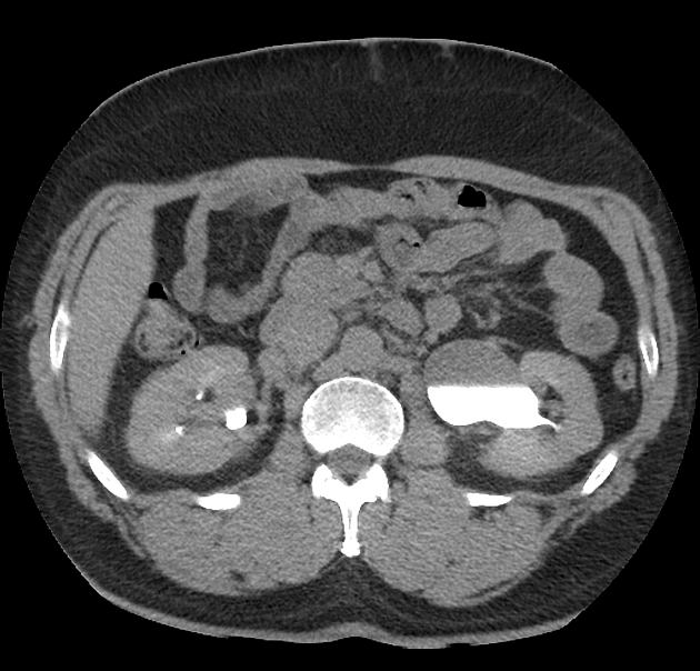
CT/MRI
a normal extrarenal pelvis will demonstrate normal renal cortical thickness, bilateral symmetrical contrast excretion, and normal-appearing calyces 3
Differential diagnosis
hydronephrosis, from whatever cause
Practical points
After evaluation with ultrasound and/or CT/MRI, if there is still confusion about whether a dilated renal pelvis is obstructed or not, renal scintigraphy can clarify.


 Unable to process the form. Check for errors and try again.