
Forbidden areas in mammography
Citation, DOI, disclosures and article data
At the time the article was created Stefano Pacifici had no recorded disclosures.
View Stefano Pacifici's current disclosuresAt the time the article was last revised Andrew Murphy had no financial relationships to ineligible companies to disclose.
View Andrew Murphy's current disclosures- Forbidden areas
- Forbidden areas in a mammogram
- Check area
- Check areas in a mammogram
- Review areas in a mammogram
- Forbidden areas in mammograms
- Forbidden area in a mammogram
- Check areas in mammograms
- Check areas in mammography
- Check area in mammography
- Review area in a mammogram
- Review areas in mammography
- Review area in mammography
- Review area in mammograms
- Check areas
- Forbidden area
- Forbidden zones
- Milky Way appearance on mammography
- Review areas (mammogram)
- Review area (mammogram)
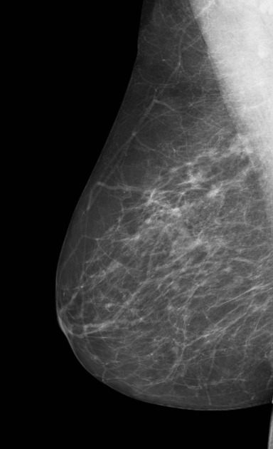
In breast imaging, forbidden, check or review areas are zones that, according to Tabár, require special attention in mammographic interpretation. These are:
- on a mediolateral oblique (MLO) view
- the "Milky Way" (retromammary fat): a 3-4 cm wide band parallel to the edge of the pectoral muscle
- retroareolar space
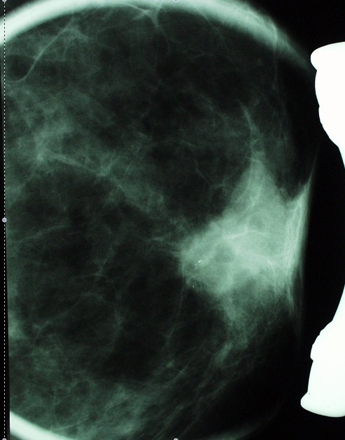
- on a craniocaudal (CC) view
- the medial half of the breast
- "no man's land": retroglandular clear space between the posterior border of the breast parenchyma and the chest wall on any view, especially on the CC view
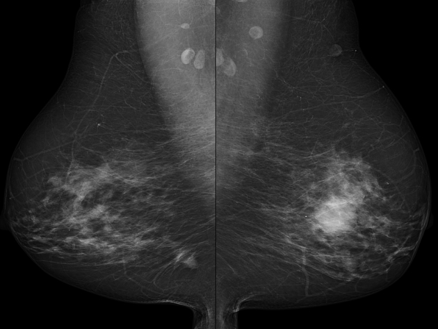
The definition "forbidden areas" is based on the incidence and distribution of cancer or de novo cancers identified in screening, demonstrating clusters located in the tail and in the inframammary fold on the MLO view, and in the tail, centrally and in the medial portions of the breast on the CC view. This warrants neutral radiographic position (no medial or lateral rotation).
References
- 1. Naylor SM, Lee L, Evans AJ. A study to find the optimal orientation of the breast for the cranio caudal view, for screening purposes. Clin Radiol. 1999;54 (12): 804-6. - Pubmed citation
- 2. Brown M, Eccles C, Wallis MG. Geographical distribution of breast cancers on the mammogram: an interval cancer database. Br J Radiol. 2001;74 (880): 317-22. Br J Radiol (full text) - Pubmed citation
- 3. Tabár L, Dean PB, Tot T. Teaching atlas of mammography. George Thieme Verlag. (2001) ISBN:0865779627. Read it at Google Books - Find it at Amazon
- 4. Hackshaw AK, Wald NJ, Michell MJ et-al. An investigation into why two-view mammography is better than one-view in breast cancer screening. Clin Radiol. 2000;55 (6): 454-8. doi:10.1053/crad.2000.0448 - Pubmed citation
- 5. Daly CA, Apthorp L, Field S. Second round cancers: how many were visible on the first round of the UK National Breast Screening Programme, three years earlier? Clin Radiol. 1998;53 (1): 25-8. - Pubmed citation
Incoming Links
Related articles: Breast imaging
- breast imaging
-
mammography
- breast screening
- breast imaging and the technologist
- forbidden (check) areas in mammography
-
mammography views
- craniocaudal view
- mediolateral oblique view
- additional (supplementary) views
- true lateral view
- lateromedial oblique view
- late mediolateral view
- step oblique views
- spot view
- double spot compression view
- magnification view
- exaggerated craniocaudal (axillary) view
- cleavage view
- tangential views
- caudocranial view
- bullseye CC view
- rolled CC view
- elevated craniocaudal projection
- caudal cranial projection
- 20° oblique projection
- inferomedial superolateral oblique projection
- Eklund technique
- normal breast imaging examples
-
mammography
- digital breast tomosynthesis
- breast ultrasound
- breast ductography
- breast MRI
- breast morphology
- breast intervention


 Unable to process the form. Check for errors and try again.