
Gastrointestinal nodular lymphoid hyperplasia is a type of nodular lymphoid hyperplasia that can be found elsewhere in the body. It is formed out of gut-associated lymphoid tissue (GALT), and most often is a diagnostic dilemma for radiologists in the stomach and terminal ileum.
On this page:
Pathology
Gut-associated lymphoid tissue (GALT) is a subset of the larger class of mucosa-associated lymphoid tissue (MALT), and it underlies the acquired immune system in the gastrointestinal tract.
GALT is formed from lymphoid aggregates in the mucosa, including lymphoid follicles and lymph nodes. Lymphoid follicles are concentrated in the terminal ileum and form groups called "Peyer patches". The epithelium above these follicles is involved with complex responses to antigens it encounters in the small bowel.
The concentration of lymphoid tissue in the terminal ileum also explains why small bowel lymphoma has a predilection for the site.
Radiographic features
Enlarged lymphoid follicles are within normal limits for young adults, which often is the same population that is evaluated for new-onset Crohn disease.
Fluoroscopy
Small bowel follow through
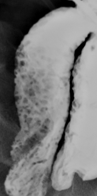
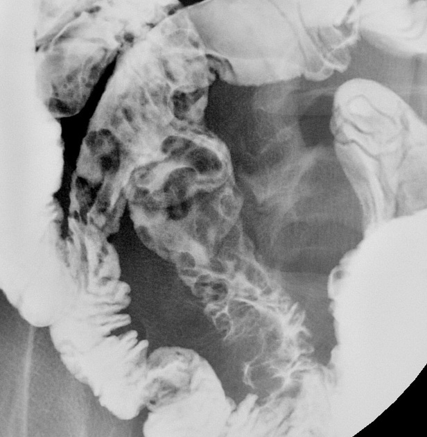
- terminal ileum
- multiple discrete, 1-3 mm nodules along the mucosal surface
- nodules should not be confluent
- small bowel folds should be normal-appearing
Differential diagnosis
- normal variant: if a few prominent nodules are found in the terminal ileum of a young person
- immunodeficiency / CVID: can be considered if a much greater than normal number of nodules are found
- lymphoma: if mucosal nodularity is larger, more confluent, and more irregular
- if small bowel folds are abnormal, one should consider


 Unable to process the form. Check for errors and try again.