Gilula three carpal arcs
Citation, DOI, disclosures and article data
At the time the article was created Umamaheswara Reddy V had no recorded disclosures.
View Umamaheswara Reddy V's current disclosuresAt the time the article was last revised Nico Behnke had no financial relationships to ineligible companies to disclose.
View Nico Behnke's current disclosures- Carpal arcs
- Three carpal arcs of Gilula
- Gilula's three carpal arcs
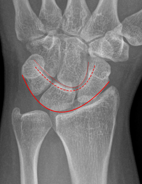
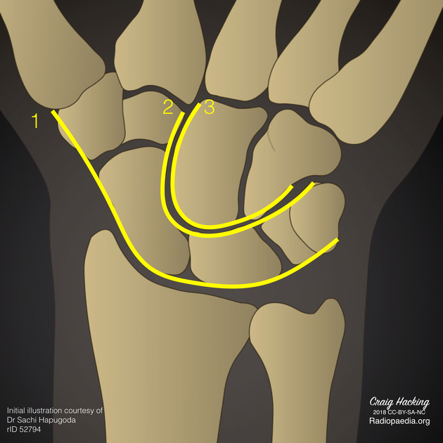
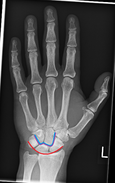
Gilula three carpal arcs are used in the assessment of the normal alignment of the carpus on PA wrist radiographs:
first arc: is a smooth curve outlining the proximal convexities of the scaphoid, lunate and triquetrum
second arc: traces the distal concave surfaces of the same bones
third arc: follows the main proximal curvatures of the capitate and hamate
Alignment
carpal bones have smooth and rounded edges to varying degrees, and lines joining these convexities form arcs; when major convexities are used in drawing
-
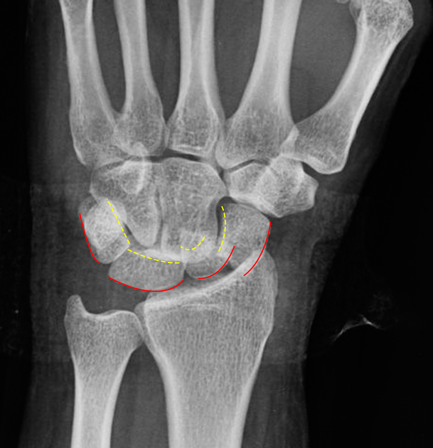
there should be no step-offs in the contour of the arcs, except for two normal variants 4
a triquetrum that is shorter in the proximal-distal dimension than the lunate creates a step-off in the first arc, but there is still a normal second arc
"bi-lobed" appearance of second carpal arc in lunate type II morphology because of a proximally prominent hamate
disrupted arc may indicate a ligamentous injury or fracture at the site of the broken arc
History and etymology
The concept of three radiographic arcs was first proposed by Louis A Gilula (1942-2014) in 1979 3,5.
References
- 1. Kaewlai R, Avery L, Asrani A, Abujudeh H, Sacknoff R, Novelline R. Multidetector CT of Carpal Injuries: Anatomy, Fractures, and Fracture-Dislocations. Radiographics. 2008;28(6):1771-84. doi:10.1148/rg.286085511 - Pubmed
- 2. Vezeridis P, Yoshioka H, Han R, Blazar P. Ulnar-Sided Wrist Pain. Part I: Anatomy and Physical Examination. Skeletal Radiol. 2009;39(8):733-45. doi:10.1007/s00256-009-0775-x - Pubmed
- 3. Gilula L. Carpal Injuries: Analytic Approach and Case Exercises. AJR Am J Roentgenol. 1979;133(3):503-17. doi:10.2214/ajr.133.3.503 - Pubmed
- 4. Loredo R, Sorge D, Garcia G. Radiographic Evaluation of the Wrist: A Vanishing Art. Semin Roentgenol. 2005;40(3):248-89. doi:10.1053/j.ro.2005.01.014 - Pubmed
- 5. Rubin D. Louis A. Gilula, MD. Radiology. 2015;274(1):308. doi:10.1148/radiol.14144047 - Pubmed
Incoming Links
- Scapholunate advanced collapse (SLAC)
- Disrupted Gilula arcs (anatomic variant)
- Bilateral pronator quadratus sign
- Scaphoid fracture
- Lunate dislocation
- Lunate dislocation
- Perilunate dislocation
- Lunate dislocation
- Gilula carpal arcs (diagram)
- 4th and 5th carpometacarpal joint dislocations
- Volar intercalated segmental instability (VISI)
- Scapholunate advanced collapse (SLAC)
- Gilula carpal arcs
- Scaphoid fracture - Mayo middle type
- Calcium pyrophosphate deposition arthropathy wrist
- Lunate avascular necrosis
Related articles: Anatomy: Upper limb
-
skeleton of the upper limb
- clavicle
- scapula
- humerus
- radius
- ulna
- hand
- accessory ossicles of the upper limb
- accessory ossicles of the shoulder
- accessory ossicles of the elbow
-
accessory ossicles of the wrist (mnemonic)
- os centrale carpi
- os epilunate
- os epitriquetrum
- os styloideum
- os hamuli proprium
- lunula
- os triangulare
- trapezium secondarium
- os paratrapezium
- os radiostyloideum (persistent radial styloid)
- joints of the upper limb
-
pectoral girdle
-
shoulder joint
- articulations
- associated structures
- joint capsule
- bursae
- ligaments
- movements
- scapulothoracic joint
-
glenohumeral joint
- arm flexion
- arm extension
- arm abduction
- arm adduction
- arm internal rotation (medial rotation)
- arm external rotation (lateral rotation)
- circumduction
- arterial supply - scapular anastomosis
- ossification centers
-
shoulder joint
-
elbow joint
- proximal radioulnar joint
- ligaments
- associated structures
- movements
- alignment
- arterial supply - elbow anastomosis
- development
-
wrist joint
- articulations
-
ligaments
- intrinsic ligaments
- extrinsic ligaments
- radioscaphoid ligament
- dorsal intercarpal ligament
- dorsal radiotriquetral ligament
- dorsal radioulnar ligament
- volar radioulnar ligament
- radioscaphocapitate ligament
- long radiolunate ligament
- Vickers ligament
- short radiolunate ligament
- ulnolunate ligament
- ulnotriquetral ligament
- ulnocapitate ligament
- ulnar collateral ligament
- associated structures
- extensor retinaculum
- flexor retinaculum
- joint capsule
- movements
- alignment
- ossification centers
-
hand joints
- articulations
- carpometacarpal joint
-
metacarpophalangeal joints
- palmar ligament (plate)
- collateral ligament
-
interphalangeal joints
- palmar ligament (plate)
- collateral ligament
- movements
- ossification centers
- articulations
-
pectoral girdle
- spaces of the upper limb
- muscles of the upper limb
- shoulder girdle
- anterior compartment of the arm
- posterior compartment of the arm
-
anterior compartment of the forearm
- superficial
- intermediate
- deep
-
posterior compartment of the forearm (extensors)
- superficial
- deep
- muscles of the hand
-
accessory muscles
- elbow
- volar wrist midline
- palmaris longus profundus
- aberrant palmaris longus
- volar wrist radial-side
- accessory flexor digitorum superficialis indicis
- flexor indicis profundus
- flexor carpi radialis vel profundus
- accessory head of the flexor pollicis longus (Gantzer muscle, common)
- volar wrist ulnar-side
- dorsal wrist
- blood supply to the upper limb
-
arteries
- subclavian artery (mnemonic)
- axillary artery
- brachial artery (proximal portion)
- ulnar artery
- radial artery
- veins
-
arteries
- innervation of the upper limb
- intercostobrachial nerve
-
brachial plexus (mnemonic)
- branches from the roots
- branches from the trunks
- branches from the cords
- lateral cord
- posterior cord
- medial cord
- terminal branches
- lymphatic drainage of the upper limb
Related articles: Wrist pathology
- alignment
- wrist fractures and dislocations
- distal radial fracture
- pediatric
- carpal bones
- Mayfield classification of carpal instability
- carpal instability
- osteonecrosis
- triangular fibrocartilaginous complex (TFCC) injuries
- ulnar-sided wrist impaction and impingement syndromes
- soft tissue and tendons
- arthritides


 Unable to process the form. Check for errors and try again.