The glossopharyngeal nerve is the ninth cranial nerve (CN IX). It exits the brainstem out from the sides of the upper medulla, just rostral to the vagus nerve and has sensory, motor, and autonomic components (TA: nervus glossopharyngeus or nervus cranialis IX).
On this page:
Images:
Gross anatomy
Origin
There are four cranial nerve nuclei in the lower pons and medulla that contribute to the glossopharyngeal nerve:
-
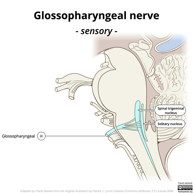
The sensory ganglion cells lie in the superior and inferior ganglia of the nerve, whose central processes pass to two nuclei:
nucleus of the tractus solitarius, located in the dorsolateral medulla oblongata and lower pons, which conveys taste sensation from the posterior 3rd of the tongue and vallate papillae
spinal nucleus of the trigeminal nerve, located in the caudal aspect of the pons, which conveys somatic sensation
The motor fibers originate in the upper part of the nucleus ambiguus in the rostral pons which receives bilateral supranuclear innervation from the corticobulbar fibers. This nucleus supplies the stylopharyngeus muscle.
The autonomic parasympathetic fibers arise in the inferior salivary nucleus located in the dorsal pons. These fibers are carried in the lesser petrosal nerve via the tympanic branch to the otic ganglion. The postganglionic fibers are distributed to the parotid gland via the auriculotemporal nerve.
Course
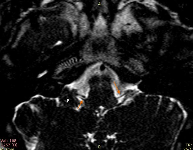
The glossopharyngeal nerve exits the medulla oblongata from the postolivary sulcus, passes laterally across the flocculus, and leaves the skull through the pars nervosa of the jugular foramen in a separate sheath of the dura mater. It then passes between the internal jugular vein and internal carotid artery. It descends in front of the latter vessel, and beneath the styloid process and the muscles connected with it, to the lower border of the stylopharyngeus muscle. It then curves forward, forming an arch on the side of the neck and lying on the stylopharyngeus and middle pharyngeal constrictor muscle. From there, it passes under a cover of the hyoglossus muscle, and is finally distributed to the palatine tonsil, the mucous membrane of the fauces and base of the tongue, and the mucous glands of the mouth.
Glossopharyngeal ganglia
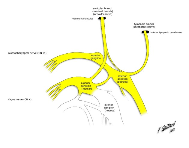
In passing through the jugular foramen, the nerve presents two ganglia, the superior and the petrous:
superior ganglion (jugular ganglion): situated in the upper part of the groove in which the nerve is lodged during its passage through the jugular foramen; it is very small and is usually regarded as a detached portion of the petrous ganglion
inferior ganglion (petrous ganglion): larger than the superior and is situated in a depression in the lower border of the petrous portion of the temporal bone called the petrosal fossula
Branches
tympanic nerve (nerve of Jacobson): the tympanic nerve exits the jugular foramen and passes by the inferior glossopharyngeal ganglion; it re-enters the skull through the inferior tympanic canaliculus and reaches the tympanic cavity where it forms a plexus (the tympanic plexus) in the middle ear cavity; the nerve travels from this plexus through a canal and out into the middle cranial fossa adjacent to the exit of the greater petrosal nerve; here the nerve becomes the lesser petrosal nerve; the lesser petrosal nerve exits the cranium via the foramen ovale and synapses in the otic ganglion. It supplies the inner surface of the tympanic membrane (the external surface is supplied by the auriculotemporal nerve and the greater auricular nerve).
muscular branch: innervates the stylopharyngeus muscle
carotid branches: (superior and inferior caroticotympanic nerves) descend along the trunk of the internal carotid artery as far as its origin, communicating with the pharyngeal branch of the vagus, and with branches of the sympathetic nerves
pharyngeal branches: are three or four filaments which unite, opposite the middle pharyngeal constrictor with the pharyngeal branches of the vagus and sympathetic nerves, to form the pharyngeal plexus: branches from this plexus perforate the muscular coat of the pharynx and supply its muscles and mucous membrane
tonsillar branches: supply the palatine tonsil, forming around it a plexus from which filaments are distributed to the soft palate and fauces, where they communicate with the greater and lesser palatine nerves
lingual branches: are two in number; one supplies the vallate papillae and the mucous membrane covering the base of the tongue; the other supplies the mucous membrane and follicular glands of the posterior part of the tongue, and communicates with the lingual nerve
carotid sinus nerve, or "Hering nerve": the branch of the glossopharyngeal nerve to the carotid sinus is the nerve that runs downwards anterior to the internal carotid artery, communicates with the vagus nerve and sympathetic trunk, then divides in the angle of bifurcation of the common carotid artery to supply the carotid body and carotid sinus; it carries impulses from the baroreceptors in the carotid sinus (concerned with blood pressure regulation) and from chemoreceptors in the carotid body (concerned with O2 pressure monitoring; however, as of 2022, there is ongoing research as to its role in metabolic control)
Supply
There are a number of functions of the glossopharyngeal nerve:
receives general sensory fibers from the tonsils, the pharynx, the middle ear, and the posterior one-third of the tongue
receives special sensory fibers (taste) from the posterior one-third of the tongue
receives visceral sensory fibers from the carotid bodies
supplies parasympathetic fibers to the parotid gland via the otic ganglion
supplies motor fibers to stylopharyngeus muscle, the only motor component of this cranial nerve
contributes to the pharyngeal plexus
Related pathology
absent gag reflex in patients with damage to the glossopharyngeal nerve: it is responsible for the afferent limb of the reflex


 Unable to process the form. Check for errors and try again.