
Presentation
Review of normal anatomy of the cranial nerves
Patient Data
Age: 25 years
Gender: Male
From the case:
Normal cranial nerves


Download
Info
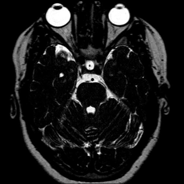
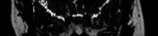
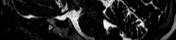
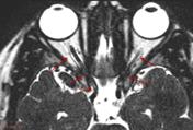
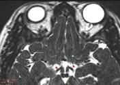
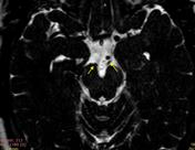
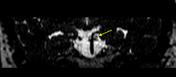
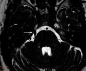
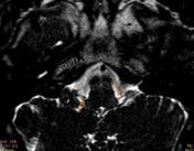
Normal MRI brain shows the different cisternal portion of the cranial nerves using SSFP with 1.4 mm thickness
From the case:
Normal cranial nerves


Download
Info
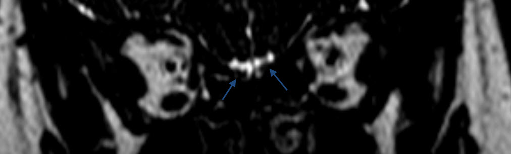
- Blue arrows refer to the Olfactory nerves (CN I) inferior to the Gyrus rectus GR.
- Red arrows refer to the Optic nerves (CN II) with the surrounding CSF in its intraocular, intracanalicular and intracranial segments. Red arrowheads refer to optic tracts and dashed red arrow to optic chiasm.
- Yellow arrows refer to Oculomotor nerve (CN III) while emerging from the medial aspect of cerebral peduncle and in-between the posterior cerebral artery and superior cerebellar artery in coronal image.
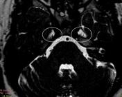
- White arrow refers to the Trigeminal nerve (CN V) and white circle refers to Meckel's cave where Gasserian ganglion located.
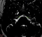
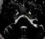
- Green arrows refer to Abducens nerve (CN VI) and green circle indicates the Dorello's canal.
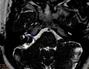
- Purple arrow refers to the Cochlear nerve (CN VIII) entering the cochlea (purple arrowhead).
- Grey arrow refers to the inferior vestibular nerve (CN VIII).
- Orange arrow refers to the Glossopharyngeal nerve (CN IX) in relation to the cerebellar flocculus F.
Case Discussion
Cranial nerves are very thin and hard to distinguish on conventional MR images. Hence the development of steady state free precession SSFP which with thin section acquisition provides high contrast resolution between CSF and other structures.


 Unable to process the form. Check for errors and try again.