
Hemotympanum is the presence of blood in the middle ear cavity. It is usually secondary to trauma.
On this page:
Clinical presentation
Typically on otoscopy a bulging red to purple to dark blue colored tympanic membrane is visible, color varying with age of the hemorrhage.
Pathology
The hemorrhage has usually bled from superficial branches of the external carotid artery, which provide the rich vasculature of the middle ear.
Etiology
- blunt trauma of the temporal bone
- temporal bone fractures
- iatrogenic nasal packing and/or epistaxis on background of an incompetent Eustachian tube
- blood dyscrasias, e.g. idiopathic thrombocytopenic purpura
- leukemia 3
- barotrauma 3
- scuba diving, air travel
- anticoagulation therapy (rare) 2
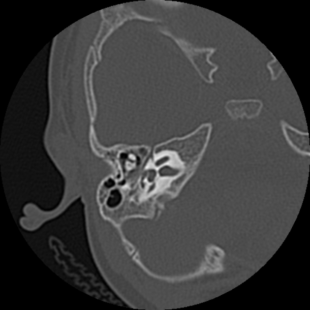
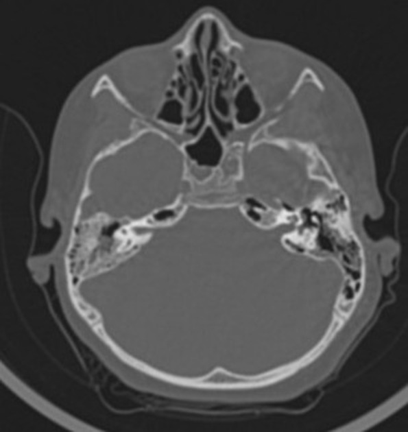
Radiographic features
On CT or MRI there is evidence of opacification of the middle ear cleft by fluid/blood products.
Treatment and prognosis
Conservative management usually suffices. Treatment of the underlying cause may also be required.


 Unable to process the form. Check for errors and try again.