Citation, DOI, disclosures and article data
Citation:
Dawes L, Silverstone L, Walizai T, et al. Hemithorax white-out (differential). Reference article, Radiopaedia.org (Accessed on 29 Mar 2025) https://doi.org/10.53347/rID-1439
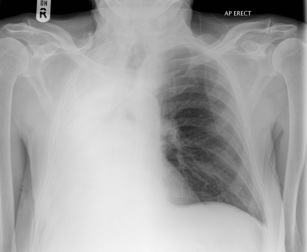
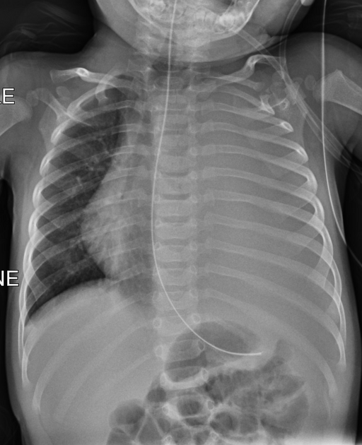
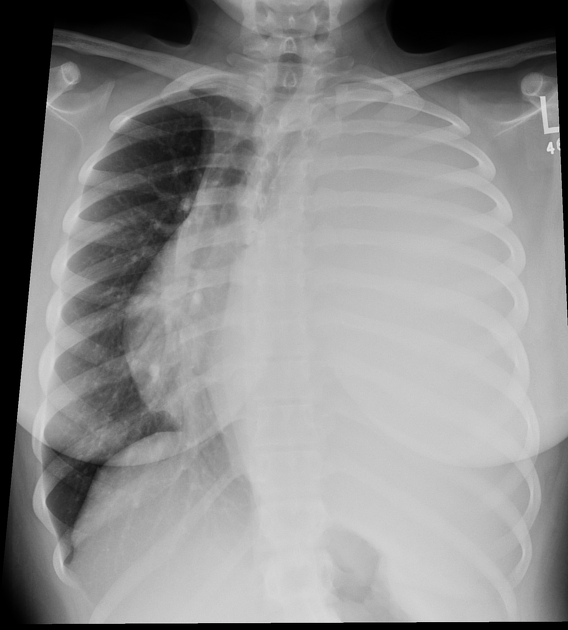
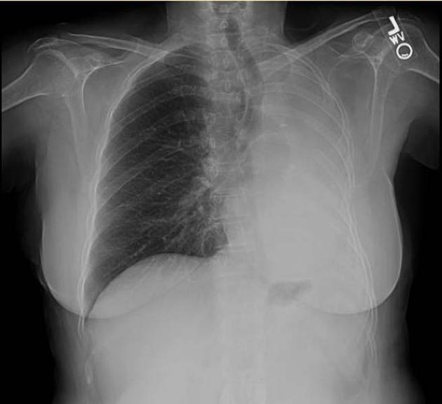
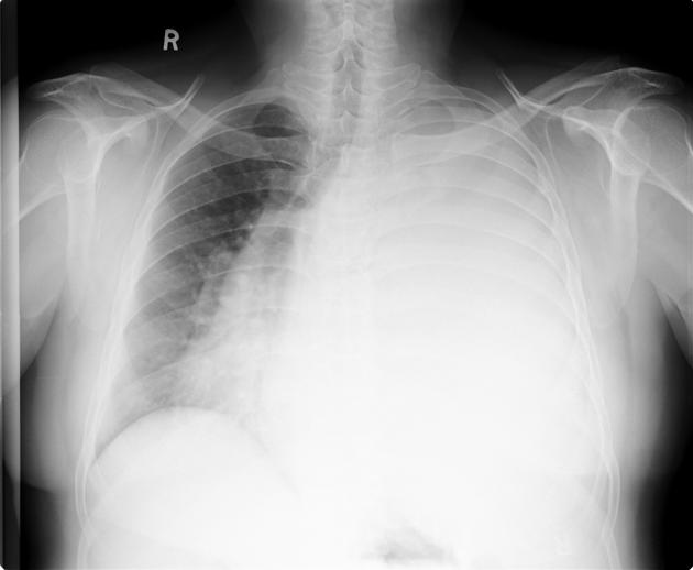
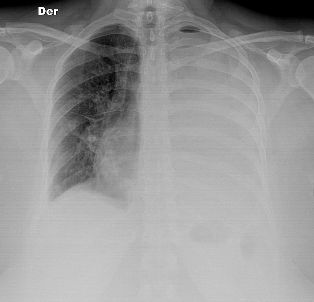
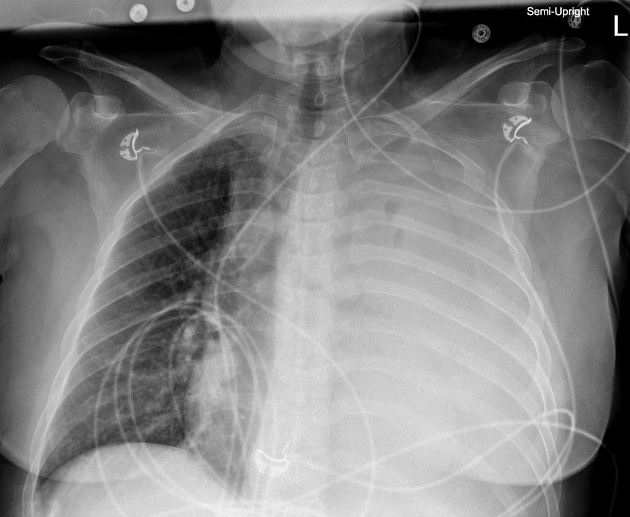
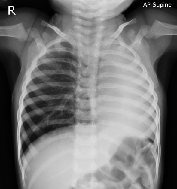
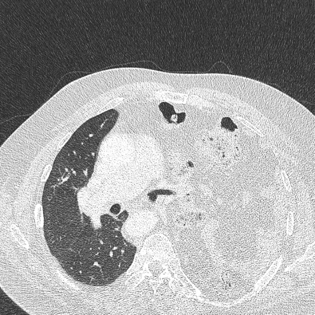
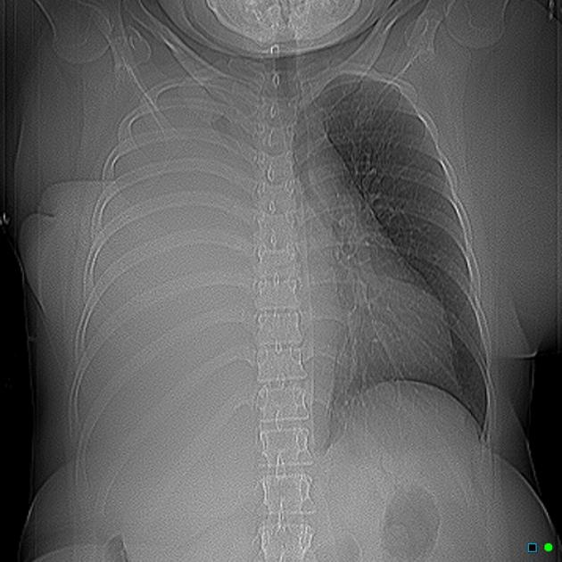
Complete white-out of a hemithorax on the chest x-ray has a limited number of causes. The differential diagnosis can be shortened further with one simple observation: the position of the trachea, heart and mediastinum. Are they central, or are they displaced towards or away from the opacified lung? Changes in volume of the opacified hemithorax will also affect rib spacing.
Volume loss
No change in volume
Increased volume
See also
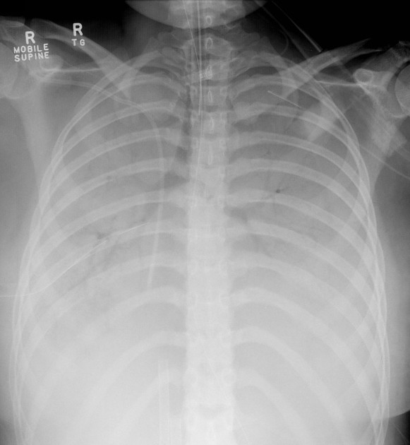
What is the most likely reason for complete opacification of this patient's left hemithorax?

-
1. Khan AN, Al-jahdali H, Al-ghanem S et-al. Reading chest radiographs in the critically ill (Part II): Radiography of lung pathologies common in the ICU patient. Ann Thorac Med. 2009;4 (3): 149-57. doi:10.4103/1817-1737.53349 - Free text at pubmed - Pubmed citation
-
2. Corne J, Carroll M, Delany D et-al. Chest x-ray made easy. Churchill Livingstone. (2002) ISBN:0443070083. Read it at Google Books - Find it at Amazon
-
3. Chapman S, Nakielny R. Aids to radiological differential diagnosis. Saunders Ltd. (2003) ISBN:0702026506. Read it at Google Books - Find it at Amazon
-
4. Susie J. Goodwin, Elise Randle, Akane Iguchi, Katherine Brown, Aparna Hoskote, Alistair D. Calder. Chest computed tomography in children undergoing extra-corporeal membrane oxygenation: a 9-year single-centre experience. (2014) Pediatric Radiology. 44 (6): 750. doi:10.1007/s00247-014-2878-3 - Pubmed
-
5. Alex M. Barnacle, Liz C. Smith, Melanie P. Hiorns. The Role of Imaging During Extracorporeal Membrane Oxygenation in Pediatric Respiratory Failure. (2012) American Journal of Roentgenology. 186 (1): 58-66. doi:10.2214/AJR.04.1672 - Pubmed
Multiple choice questions:
Promoted articles (advertising)


 Unable to process the form. Check for errors and try again.